Researchers pinpoint area of brain linked to bipolar disorder
 A volume decrease in specific parts of the brain’s hippocampus – long identified as a hub of mood and memory processing – was linked to bipolar disorder in a study led by researchers at McGovern Medical School. The research was published in Molecular Psychiatry.
“Our study is one of the first to locate possible damage of bipolar disorder in specific subfields within the hippocampus,” said Bo Cao, Ph.D., first and corresponding author and a postdoctoral fellow in the Department of Psychiatry and Behavioral Sciences at McGovern Medical School. “This is something that researchers have been trying to answer. The theory was that different subfields of the hippocampus may have different functions and may be affected differently in different mood disorders, such as bipolar disorder and major depression disorder.”
Cao hopes the study, which was funded in part by the National Institute of Mental Health (NIMH), will pioneer future research on details within the hippocampus as a marker for precise diagnosis and positive treatment response of bipolar disorder.
Approximately 6 million Americans suffer from bipolar disorder. Bipolar I disorder is characterized by mood changes that can swing from a high-energy, manic state to a low-energy, depressive state. The disorder can affect sleep, energy level and the ability to think clearly, according to the National Institutes of Health. It can interfere with a person’s ability to work and perform daily living activities, and could lead to suicide attempts. Patients with bipolar II disorder do not experience the full-blown manic episodes, but may have a less severe high-energy state.
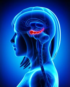
The research team used a combination of magnetic resonance imaging (MRI) and a state-of-the-art segmentation approach to discover differences in the volumes of subfields of the hippocampus, a seahorse-shaped region in the brain. Subjects with bipolar disorder were compared to healthy subjects and subjects with major depressive disorder.
Researchers found that subjects with bipolar disorder had reduced volumes in subfield 4 of the cornu ammonis (CA), two cellular layers and the tail portion of hippocampus. The reduction was more severe in patients with bipolar I disorder than other mood disorders investigated.
Further, in patients with bipolar I disorder, the volumes of certain areas such as the right CA 1 decreased as the illness duration increased. Volumes of other CA areas and hippocampal tail were more reduced in subjects who had more manic episodes.
Jair Soares, M.D., Ph.D., professor, chairman and the Pat R. Rutherford, Jr., Chair in the Department of Psychiatry and Behavioral Sciences at McGovern Medical School, was senior author of the paper. Other McGovern Medical School co-authors included Benson Mwangi, Ph.D.; Henrique Amaral-Silva, Ph.D.; Jonika Tannous; Mon-Ju Wu, Ph.D.; and Giovana Zunta-Soares, M.D. Soares is a member and Tannous is a student of The University of Texas Graduate School of Biomedical Sciences at Houston.
Funding from the NIMH, part of the National Institutes of Health, was under grant R01 085666. Additional funding came from the John S. Dunn Foundation and the Pat Rutherford, Jr. Endowed Chair in Psychiatry through Soares.
-Deborah Mann Lake, Office of Public Affairs