Projects
An area of major interest is to determine the stiffness of a sample (Elastic Modulus). We can evaluate the effects of several physiological processes by measuring the elastic response (Young’s modulus) on cells and tissues. For example, the elasticity of the cell membrane can vary between cell types as a function of growth, differentiation, disease or treatment. We are now able to evaluate the effects of treatment in cancer cells, analyze the efficiency of skin healing, or simply observe the changes in DNA structures, all at the nano-metric scale.
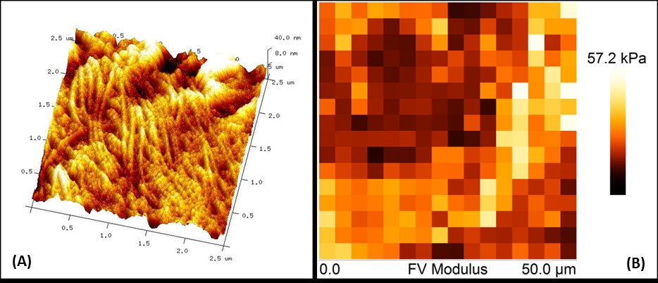
Additionally, we can map the distribution of elastic responses on the sample surface (force volume maps) by combining force curve measurements with topographic imaging, as shown in the Figure 1 with tissue samples. Figures 1 through 3 represent a few of the applications afforded by our AFM laboratory. The unique properties of Atomic Force Microscopy offer unlimited applications in medical research. This technology can provide valuable insights about the mechanisms of a disease and assess a successful treatment pathway for its cure.

Figure 1. Pictured are the elastic properties associated with skin healing. Color-coded force maps were generated over skin biopsies to determine collagen deposition as a promoter of wound healing in transgenic mice. (A) AFM micrograph obtained at 2.5μm (x-y) showing topography of a skin wound. (B) Force Volume map of a 50μm (x-y) scan of the same skin area.

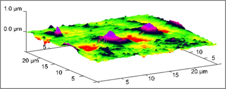
Figure 2. Liposome endocytosis occurring at 60min of incubation in HCAEC’s. 3D-AFM micrograph obtained at 25μm (x-y) illustrating the engulfment of liposomes conjugated with gold nanoparticles during membrane internalization.

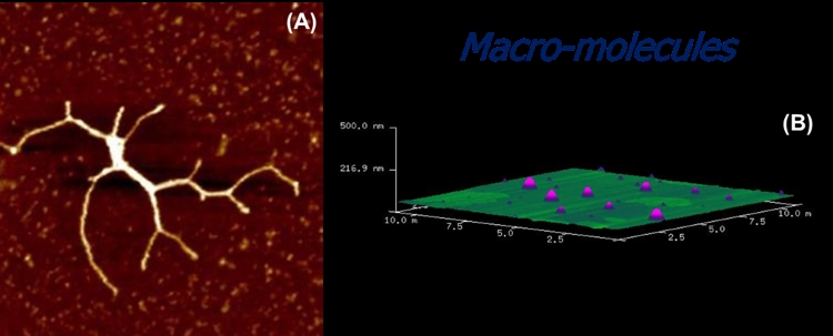
Figure 3. (A) AFM image of a full-length myosin protein visualized at RT° on mica. Scan to 2μm (x-y) using tapping mode in air. (B) 3D-AFM micrograph of DPPC liposomes deposited on mica.