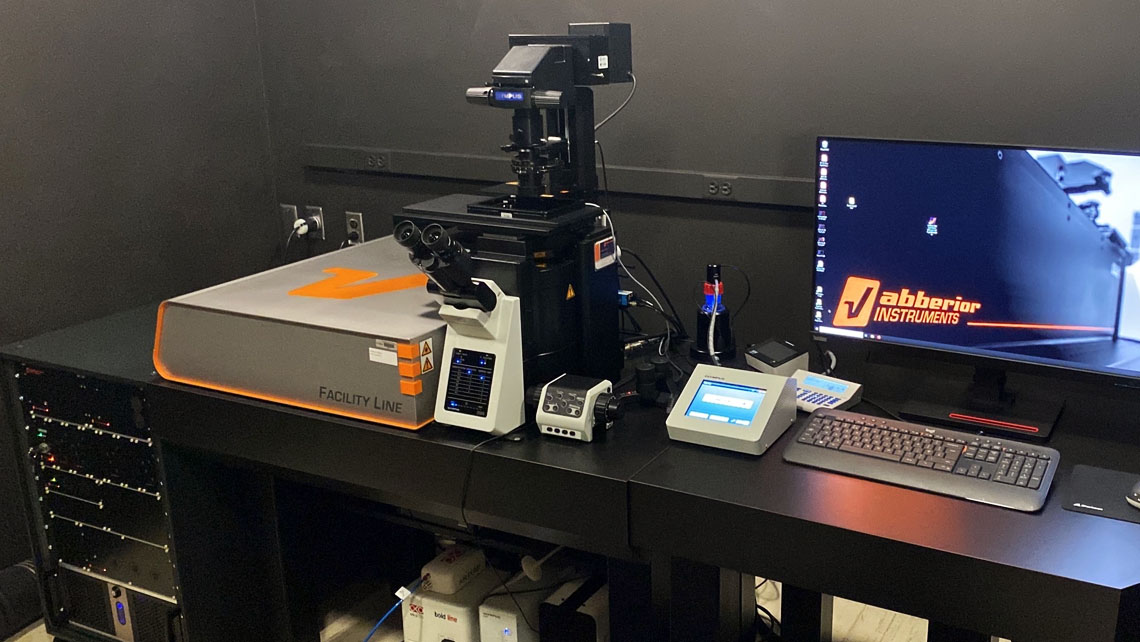
Abberior Facility Line 3D Stimulated Emission Depletion (STED) Microscopy

Text

Key features include:

4 Excitation and 1 Depletion Laser Lines

Excitation Lasers

  • 405 nm, 20 mW
  • 485 nm, 1 mW pulsed@ 40 MHz
  • 561 nm, 200 µW pulsed @ 40MHz
  • 640 nm, ~1 mW pulsed @ 40 MHz

Depletion Laser

  • 775nm, > 2750mW @40Mhz

Objectives

  • 10x Air / NA 0.4, WD: 3.1 mm
  • 40x Air / NA 0.95, WD: 0.18 mm
  • 60x Oil / NA: 1.42, WD: 0.15 mm
  • 60x Silicone Oil / NA 1.3, WD: 0.3 mm
  • 60x Water / NA 1.2, WD: 0.28 mm
 

  • Dual MATRIX Spectral Detectors: Enables significant reduction of background levels on the basis of physics – no estimation procedure such as deconvolution. MATRIX detects the background for each pixel in the STED and confocal image and allows to subtract it (“differential detection”). The final image is greatly suited for further processing and analysis, e.g., deconvolution, particle counting, etc.

  • FLEXPOSURE – Adaptive Illumination: Analysis of (live) Cellular Structures with Unprecedented Detail and Signal-to-Noise: RESCue imaging for light dose reduction with confocal (one-photon or two-photon excitation) and STED Light dose minimization by up to two orders of magnitude to leverage live cell imaging. Bleaching reduction applicable to all fluorescent markers. Based on concurrent, pixel-wise decision-making of whether structure is present in the current pixel.

  • RAYSHAPE – Adaptive Optics System: Deformable mirror-based correction of arbitrary aberrations for all beams. More than 140 elements with a stroke of several microns ensure precise correction, even of high-order aberrations. Compensates for sample-induced aberrations, e.g. when focusing into a refractive-index mismatched sample, or into samples with internal inhomogeneities. Applied spherical correction changes with focus to ensure automatic correction at all focal depths.