Equipment
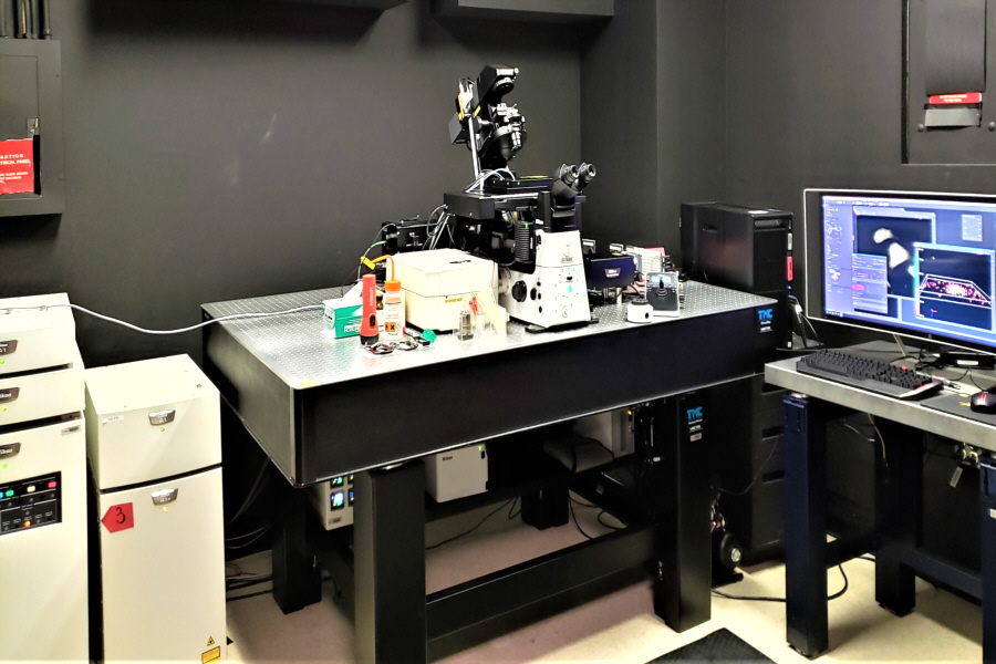
Nikon N-STORM Super-Resolution Microscope System with TIRF |
|
 |
|
| This new Nikon n-STORM Super-Resolution system is tailored toward single molecule imaging using Stochastic Optical Reconstruction Microscopy (STORM) and/or PhotoActivated Localization Microscopy (PALM) with high accuracy localization to create images with spatial resolution up to 20nm (10-fold over conventional optical microscopy) and is capable of multi-color imaging in both 2D and 3D modes. Total Internal Reflection Fluorescence (TIRF) microscopy capabilities. | |
Key features include: |
|
6 laser lines |
Objectives |
|
|
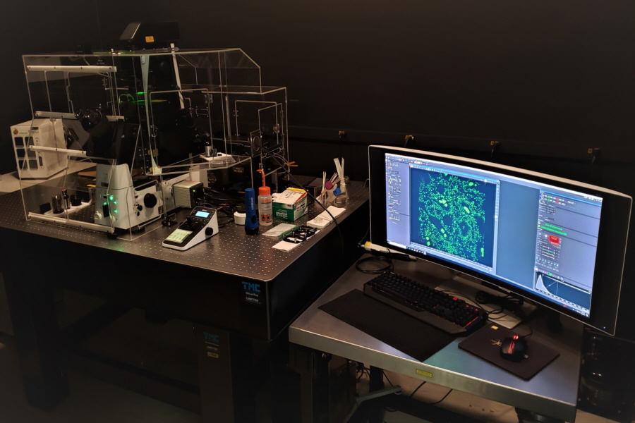
Nikon N-SIM Structured Illumination Super-Resolution Microscope System |
|
 |
|
| The system uses Structured Illumination technology, which gives a two-fold enhancement in resolution (over conventional optical microscopy). Has ability to image dynamic events in live samples in both 2D, 3D and TIRF-SIM (higher signal-to-noise to enhance resolution even further). Includes top of the line super-resolution and TIRF objectives (100X) have superior optics with the least amount of lens defects/aberrations. A wide-ranging illumination and detection systems to give users greater flexibility. |
|
Key features include: |
|
| 6 laser lines | Objectives |
|
|
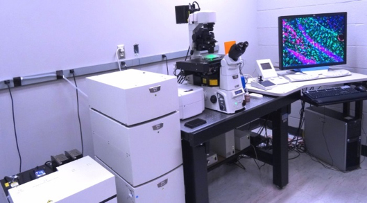
Nikon A1R Confocal Laser Microscope System |
|
 |
|
| The Nikon A1R is a modular confocal system consisting of a separate scan head, standard 4 PMT detector module plus an additional PMT for pseudo-DIC imaging, control module, laser module, and spectral detector module. The A1R has a standard high-resolution confocal mode up to 4096 x 4096 pixels as well as a high-speed resonant mode capable of scanning 230 frames per sec at 512 x 64 pixels. In addition to laser-based, confocal image acquisition, this Nikon platform also utilizes a high-end digital camera for non-confocal image acquisition.
The updated A1R has a new multi-detector unit with 2 GaAsp PMT detectors to give brighter signals, enhanced signal-to-noise ratio, and less phototoxicity. The updated A1R has the ability to perform Enhanced Resolution algorithms on point scanning confocal datasets to further increase resolution of images, and also Retirement/Replacement of Confocal Workstations allows for future expansion and faster processing, including GPU 3D Rendering.
|
|
Key features include: |
|
| 6 laser lines | Objectives |
|
|
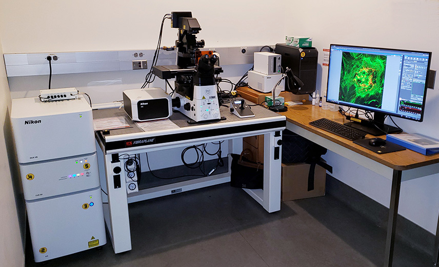
Nikon AX-R Confocal Laser Microscope System @ IMM |
|
 |
|
| Located in the Center for Advanced Microscopy Annex at the Institute for Molecular Medicine
The new generation AXR is a modular confocal system containing Ti2-E microscope, four laser unit, scan head, and detector module with two tunable detectors for green and red and two detectors with filters for blue and far red. The system has a spectral detector module and additional PMT for pseudo-DIC imaging. Control module manages the microscope, motorized stage and peripherals. This Nikon platform also utilizes a high-end digital Color camera Fi3 for non-confocal image acquisition.AXR has two confocal modes: Resonant (high speed) and standard Galvano (slower, capable of scanning 8192 x 8192 pixels). The Resonant scanning minimizes disturbance to samples, and has large field of view 25 mm and band of scanning options with resolution up to 2048 x 2048 pixels. Setup of illumination, detectors, and bandpasses for both modes is done logically and automatically. |
|
Key features include: |
|
| 4 laser lines | Objectives |
|
|
Nikon A1 Confocal Laser Microscope System + PicoQuant |
|
 |
|
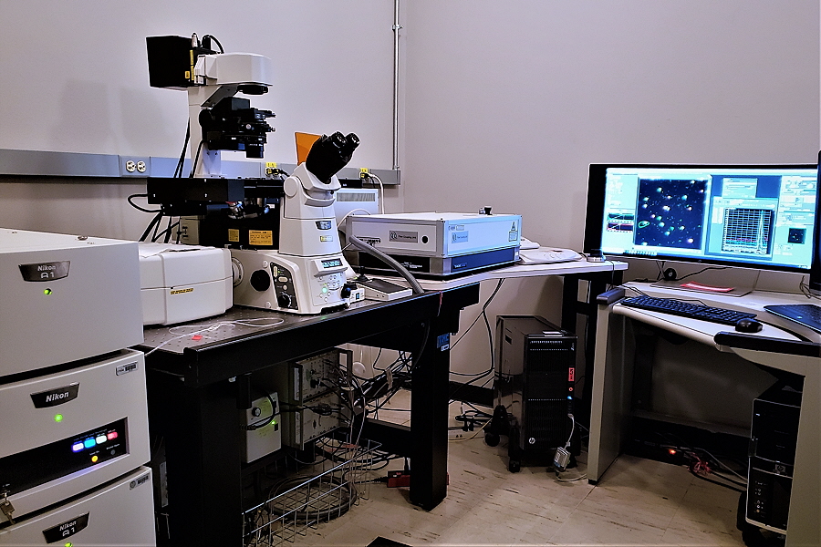
| The Nikon A1 is a basic confocal system consisting of a separate scan head, standard 5 PMT detectors, control module, and laser module. The A1 has a new multi-detector unit with 2 GaAsp PMT detectors to give brighter signals, enhanced signal-to-noise, and allow less phototoxicity. High-resolution confocal mode permits up to 4096 X 4096 pixels. This laser scanning confocal system is similar to that of the Nikon A1R in that it is configured on the Nikon TiE Widefield, inverted, fluorescence microscope. NIS Elements imaging software is used to control acquisition and analysis.
The PicoQuant Imaging System is an upgrade package added to the Nikon A1 system for the purpose of performing Fluorescence Lifetime Imaging (FLIM). This system employs pulsed lasers for the purpose of assessing protein-protein interactions and is unique to the IBP Imaging Core. The system is equipped with 439 nm (121 mW peak power) and 483 nm (136 Mw peak power) lasers.
|
|
Key features include: |
|
| 4 laser lines | Objectives |
|
|
Lambert LIFA Fluorescence Lifetime Imaging Microscopy System |
|
 |
|
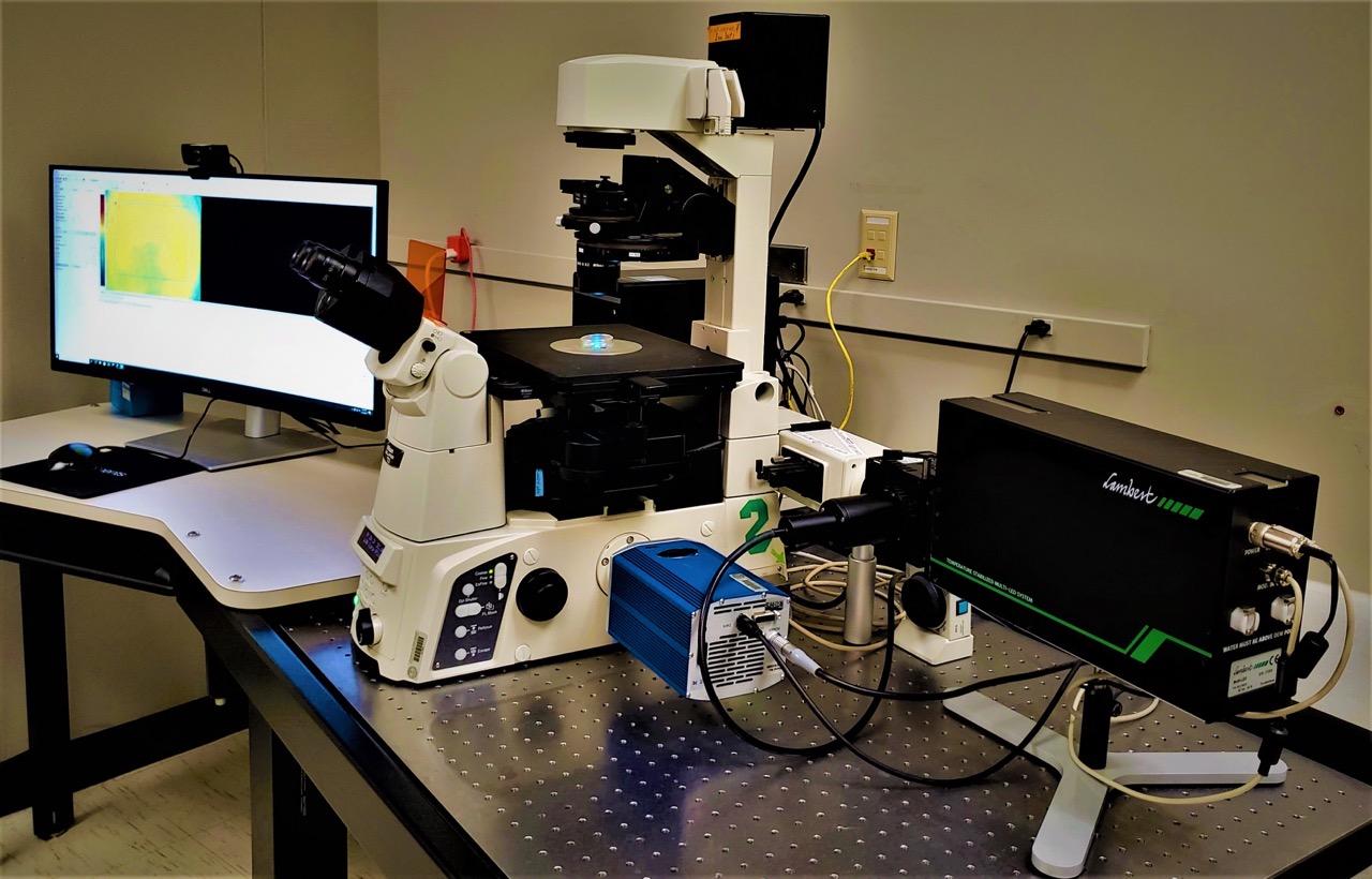
| The Lambert FLIM system is a fully developed system for frequency domain measurement of fluorescence lifetime with high accuracy, stability and acquisition/processing speed.
|
|
Key features include: |
|
| Nikon TiE Widefield fluorescence microscope | |
| High power multi-LED unit with peak emissions at: | Objectives |
|
|
Calcium Rig |
|
 |
|
| The Nikon TiE Imaging System is a high resolution system designed for fluorescence imaging of small molecules in living cells. It is a filter-based, wide-field fluorescence system designed for measurement of intracellular signaling molecules including calcium ions, hydrogen ions (pH), nitric oxide, and other small molecules using appropriate fluorophores. Nikon’s NIS Elements imaging software is used to control acquisition and analysis of captured images. The software features include capabilities for single wavelength, multiple wavelength, and ratiometric mode fluorescence imaging.
|
|
Key features include: |
|
| 6 laser lines | Objectives |
|
|
IVIS LUMINA |
|
 |
|
| The IVIS Lumina XR is live, whole animal imaging system capable of performing longitudinal fluorescence, bioluminescence and X-ray scans on in vivo and ex vivo subjects. The IVIS Lumina XR is capable of imaging all common fluorescent and bioluminescent reporters/dyes ranging from green (~488 nm) to near-infrared. The unit also contains an isoflurane-based, gas anesthesia system used for live animal studies, though investigators may utilize other methods of anesthesia provided they are approved and documented by the UT-IACUC.
|
|
Key features include: |
|
| IVIS Lumina XR | X-Ray Module |
|
|
| optical zoom lens attachment for close up and high-resolution X-ray | |
Data Processing Computers: |
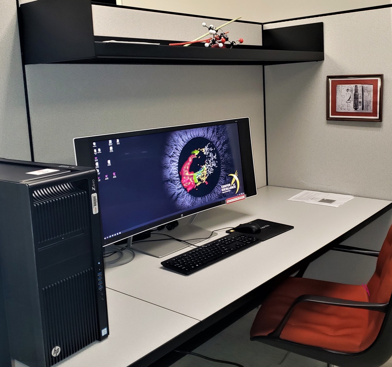
| Workstation 1: Nikon Advanced Super-Resolution Image Analysis |
 |
| Workstation 2: General Image Analysis: |
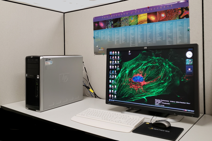 |
| NIS Elements AR; Meta Morph; LIFA-FLIM; Living Image; Image J; Slidebook; AutoQuant, and Adobe Photoshop elements 2.0. |