Craniofacial Malformations
Congenital Facial Anomalies
Congenital deformities affect the face and the upper and lower jaws. This includes children who have craniofacial deformities such as Apert’s, Crouzon’s, and Treacher-Collins, Pierre Robin Complex, hemifacial microsomia, etc. Initial treatment of these children focuses on their ability to breathe and eat normally. Some of these infants will need a tracheotomy and possibly a feeding tube. The surgical treatment of children with these craniofacial deformities usually was delayed until adolescence and involved single or double jaw-correction (orthognathic surgery). This approach is still used in some patients with mild deformities. Recently, Distraction Osteogenesis has been used to treat these patients at an earlier age. Distraction osteogenesis has been used in the newborn with respiratory difficulties and small lower jaw to stretch the lower jaw and avoid the use of tracheotomy.
Craniosynostosis
Craniosynostosis is the premature closure of one or more sutures in the infant’s skull. The treatment of craniosynostosis has changed significantly over the last several decades. Initially the treatment involved removing the affected suture(s). The simple removal of affected suture(s) was replaced by more extensive reconstruction of the craniofacial skeleton. More recently a new microscopic approach to craniosynostosis has been pioneered for treatment of infants.
In older children and those who have multiple suture synostosis, extensive reconstruction of the craniofacial skeleton is utilized. This involves an incision in the scalp from ear to ear. The affected suture is removed and the surrounding bones placed in the correct position utilizing small absorbable plates and screws. Surgery usually lasts for 2-4 hours and the child is usually hospitalized from 3-5 days. Most of patients require a blood transfusion and have significant post-operative swelling. In selected patients who have craniosynostosis of the sagittal and/or coronal sutures, helmet therapy is needed after surgery.
In children who are less than 3-months of age, a new microscopic approach is employed. This involves smaller incisions in the scalp and removal of the affected suture(s) through an endoscopic approach. Surgery usually last 1-2 hours, and the child is hospitalized for only 1-2 days. Most patients do not require a blood transfusion and have minimal swelling post-operatively. All patients require helmet therapy after surgery.
Position-induced Head Shape Abnormalities
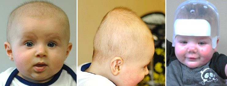
Infants with positional deformities may present with a number of different head shapes. They may have unilateral flattening of their posterior skull, which is plagiocephaly, and/or forehead bulging along with bilateral flattening, which is brachycephaly. When they have a long, narrow skull it is scaphocephaly. Deformities result from positioning during pregnancy, sleeping position, or from neck tightness. An asymmetrical skull can result in asymmetries of the face causing various functional problems that affect chewing, speech, breathing, and vision. Some of these infants can be managed with just positioning, such as changing their sleeping position. If the deformity persists, however, treatment may be necessary.
The treatment of positional deformities is with a molding cap. Ideally, the treatment is begun during the first six months of life. The band is worn 23 hours a day and requires 3-4 months of wear. Severe cases or older children require longer periods of correction. The result of wearing band is typically a 70 percent rate of improvement, but the correction rate correlates with the severity of the deformity and the age that treatment commenced. Long-term follow up in these children reveals that the correction does not relapse with continued growth.
Torticollis
Torticollis (stiff neck)- torticollis is the medical term for stiff neck. The incidence of torticollis in infants has dramatically increased since 1999 when the American Academy of Pediatrics began their “Back to the Back” campaign. Torticollis is not a diagnosis but a sign of an underlying problem. Most children suffer a stiff neck as a result of limited space in the womb and/or birth trauma. Other causes include cervical spine abnormalities, hip dysplasia, muscle spasm, ocular muscle imbalance and muscle tumor.
A child with torticollis usually presents with a head tilt. The head tilts to the side with stiff neck while the chin points away. Treatment involves repositioning and neck exercises. Most patients under a year of age respond to these conservative measures. Older patients may require surgery which involves release of the affected neck muscle. Failure to treat a stiff neck will eventually result in twisting of the child’s face.