CiCHD researchers publish groundbreaking 3D bioprinted model of early human heart development in Nature Communications
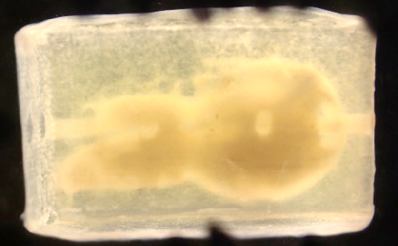
Incorporating human heart muscle cells (cardiomyocytes) into the 3D bioprinted model of the human embryonic heart tube
Researchers affiliated with the Center for Innovation in Congenital Heart Disease (CiCHD) have published a landmark study in Nature Communications introducing what is believed to be the first fully human-based, 3D bioprinted, and perfused model of the embryonic heart tube—the earliest structural stage of human heart development.
Modeling early human heart development using an iPSC-based 3D bioprinted model of embryonic heart tube (external link) represents a major advancement in the field of developmental cardiology and congenital heart disease research, providing scientists with a powerful new platform to investigate how the human heart forms and how developmental abnormalities can lead to congenital heart defects.
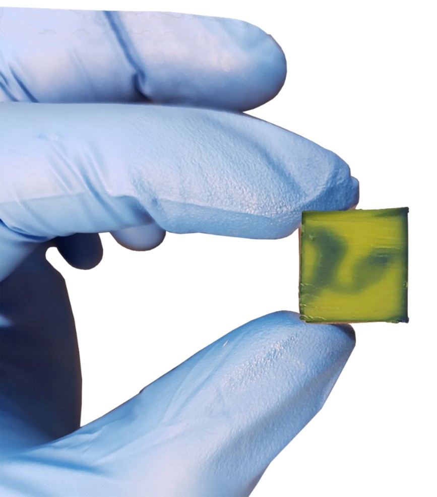
A 3D bioprinted model of the human embryonic heart tube
For decades, the earliest stages of human heart development have remained largely inaccessible to direct study. The newly developed bioengineered platform recreates key features of the embryonic heart tube using human-induced pluripotent stem cells (iPSCs), advanced 3D bioprinting technologies, and physiologically relevant perfusion systems. By mimicking the environment in which the earliest human heart structures emerge and begin functioning, the model offers an unprecedented opportunity to observe, analyze, and experimentally manipulate developmental processes that were previously beyond reach.
“Congenital heart disease is fundamentally a developmental disorder,” said CiCHD Director Vahid Serpooshan, PhD. “To understand and ultimately prevent or better treat these conditions, we need models that accurately capture the earliest events of human cardiac development. This work provides a new framework for studying those events in a controlled and reproducible way.”
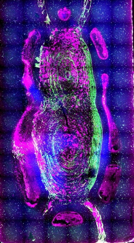
Microcopy imaging of the cross-section of bioprinted heart tube, illustrating the human cardiomyocytes (red) and endothelial cells (green) within the 3D structure
The research was led by Linqi Jin, PhD, as part of her doctoral work and reflects years of interdisciplinary collaboration spanning bioengineering, stem cell biology, and cardiovascular research. The platform enables researchers to examine developmental mechanisms, identify factors that contribute to congenital heart disease, and evaluate emerging therapeutic strategies in a human-specific system. The publication exemplifies the CiCHD mission of advancing congenital heart disease care through bioengineering and translational research. By integrating expertise in tissue engineering, regenerative medicine, and cardiovascular science, the center is developing next-generation human models that bridge the gap between laboratory discovery and clinical impact.
“This achievement demonstrates the transformative potential of combining stem cell technologies with advanced biomanufacturing approaches,” said Serpooshan. “We are moving beyond simply observing development—we are beginning to recreate it. That capability opens entirely new avenues for understanding congenital heart disease and developing personalized therapeutic solutions.”
As CiCHD continues to expand its research portfolio, innovations such as this 3D bioprinted heart development platform are expected to accelerate discoveries that improve outcomes for children and adults living with congenital heart disease.
The study (external link) is published in Nature Communications and marks another significant milestone in CiCHD’s commitment to redefining the future of congenital heart disease research and care.