Can Machine Learning Identify the Tumor Attachment Site of a Sinonasal Inverted Papilloma?
 In a retrospective review of patients with inverted papilloma (IP) who underwent resection during 2004-2021 at a tertiary care institution in the Texas Medical Center, researchers at UTHealth Houston concluded that radiomics and machine-learning applications hold great promise as novel future technologies for identifying tumor origin prospectively.
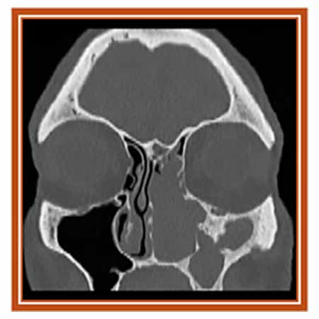
“Inverted papilloma, a benign tumor of the paranasal sinuses, is often suspected based on its appearance on CT scans – a characteristic remodeling of the bone at the attachment site,” says Martin Citardi, MD, professor and chair of the Department of Otorhinolaryngology-Head and Neck Surgery and vice dean for technology at UTHealth Houston. “Our goal in this study was to develop a machine-learning algorithm that would recognize the attachment site on a CT scan and compare the algorithm’s success with actual surgical findings. With its characteristic radiographic appearance, IP shouldn’t be a surprise diagnosis at the time of surgery done for nasal polyps, but sometimes it is. If we can identify the attachment site in advance, we can plan the surgery accordingly and prepare the patient and surgical team for either a simple or more involved procedure.”
When the research team applied the algorithm, it picked up the attachment site in 83% of revision cases, but only 62% of primary of cases. “These were good results, but the detection percentage has to be consistently above 85% to meet the threshold of acceptability,” Dr. Citardi says. “We’re working on further refining the machine-learning model to fine tune it for greater accuracy and usability.”
The research was presented by Sean McKee, MD, postgraduate year 4 resident, at the American Rhinologic Society (ARS) meeting held in Boston. The otolaryngology team worked with researchers at the McWilliams School of Biomedical Informatics and its Center for Precision Health at UTHealth Houston.
Postgraduate year 4 resident Jumah Ahmad, MD, presented the results of a second, related study in an abstract entitled “Retrospective Review of Inverted Papilloma Attachment Site Distribution with a 3D Heat Map” at the ARS meeting at COSM.
“Congruent to the literature, the most common attachment site of IP in our patients was the maxillary sinus,” Dr. Citardi says. “Contrary to the literature, the superior, posterior, and lateral maxillary walls were more commonly involved than the lateral nasal wall. We concluded that this 3D heat map of IP attachment site distribution can be insightful when reviewing patients’ preoperative imaging. A pictorial representation is a much more sophisticated way of describing location and presumably provides a truer representation of the pathology.”