Median Arcuate Ligament Syndrome (MALS)
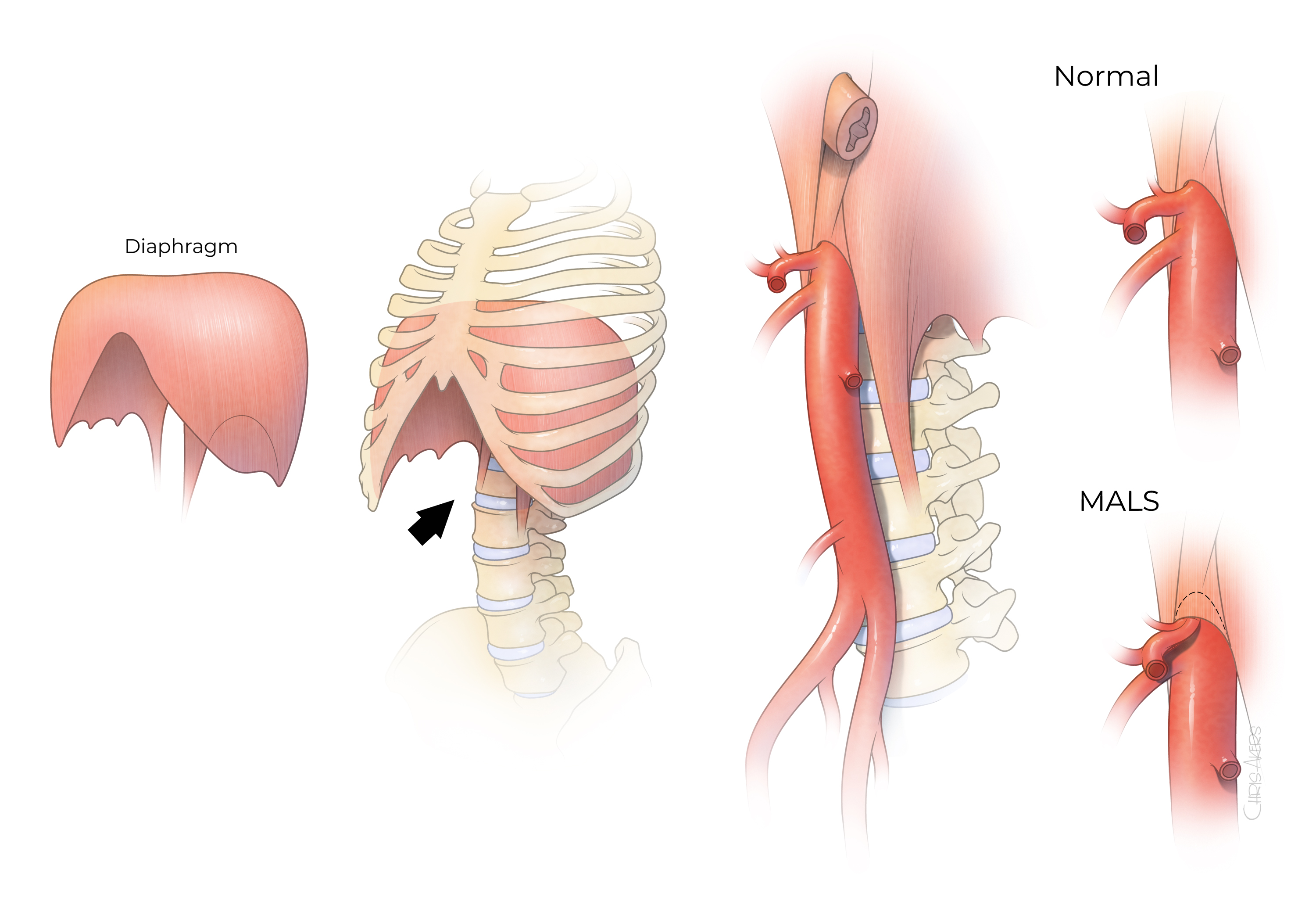
Figure 1. Illustration of the diaphragmatic muscle which separates the chest from the abdominal cavity. The aorta, the largest blood vessel that brings blood supply from the heart into all the organs, travels through the chest on top of the vertebral bodies and enters the abdomen via an opening at the diaphragmatic muscle. The median arcuate ligament is the muscle and fibrous structure that wraps around the aorta at the diaphragmatic opening. Patients with MALS have severe compression of the celiac artery, the first large branch from the aorta as it enters the abdomen. This compression is typically worse during deep expiration.
Median arcuate ligament syndrome (MALS) is an uncommon condition that predominantly affects young individuals due to excessive compression of the celiac artery, the first large branch of the abdominal aorta located near the median arcuate ligament and celiac ganglion (Figure 1). The cause remains unknown but may be due to compression of the sensitive outer arterial wall layer by the muscle and ganglionic tissue, direct irritation or inflammation of sympathetic nerves, and possibly intermittent decrease in blood supply to the upper gastrointestinal tract. The diagnosis of MALS can be difficult because most patients present with nonspecific upper abdominal pain and because compression of the artery itself is not sufficient to determine cause and effect.