Introducing the new 3D STED microscope at McGovern
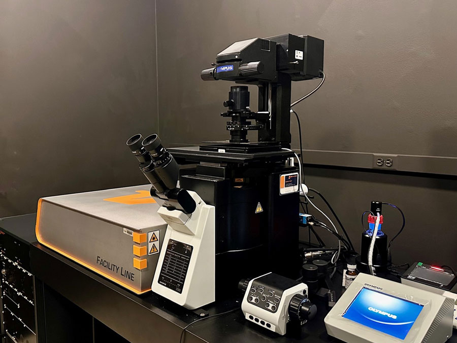
A new chapter in scientific discovery is unfolding at McGovern Medical School with the installation of a cutting-edge super-resolution microscope: the Abberior Facility Line Stimulated Emission Depletion (STED) system.
This innovative technology is the first 3D-STED microscope in the Texas Medical Center, opening unprecedented avenues for researchers to explore molecular interactions at the nanometer scale — even deep within tissue samples that are normally inaccessible to other super-resolution techniques.
“We believe this new STED microscope will transform our understanding of complex biological processes,” said Travis Moore, PhD, assistant professor in the Department of Integrative Biology and Pharmacology and director of the Center for Advanced Microscopy. “3D-STED’s ability to volumetrically image deeper into tissues and maintain ultra-high resolution provides a powerful tool to investigate tissue pathologies and signaling pathways in ways we couldn’t before.”
The Abberior Facility Line STED system comes to McGovern thanks to an NIH S10 High-end Instrumentation grant (S10 00036331) award to Moore. Housed at the Center for Advanced Microscopy, this new instrument builds on the facility’s mission of providing state-of-the-art fluorescence microscopy to investigators across multiple disciplines.
“This investment will spark new collaborations and push the boundaries of what’s possible in field like neuroscience, cellular biology, and disease pathology,” said Ryan Durham, PhD, manager of the Center for Advanced Microscopy.
What makes STED stand out compared to other super-resolution techniques such as Single-Molecule Localization Microscopy (SMLM) or Structured Illumination Microscopy (SIM)?
Unlike SMLM, which can be time-consuming and sometimes challenging to implement in thick specimens, STED can image robustly in tissue slices up to 80 microns thick at an axial resolution of 70 NM (5x scanning confocal) — whether they are paraffin-embedded or cryopreserved. And while SIM can offer improved resolution compared to diffraction-limited microscopy, STED goes a step further by routinely achieving lateral resolution down to 20 nm. This exceptional clarity allows researchers to see the molecular machinery of cells with remarkable detail, helping them to pinpoint key interactions and structural changes.
Another major advantage of the Abberior Facility Line STED system is its versatility and user-friendliness. Equipped with adaptive illumination that reduces photobleaching and a deformable mirror for correcting refractive index changes in thicker samples, the microscope provides consistent, high-quality imaging. Better still, it operates much like a traditional laser scanning confocal microscope — super-resolution capabilities can be activated at the flip of a switch.
“What really excited me about this new STED system is how effortlessly we can incorporate it into our current lab protocols,” said Ruth Heidelberger, MD, PhD, Frederic B. Asche Char in Ophthalmology and professor in the Department of Neurobiology and Anatomy. “By simply switching our fluorescent tags to STED-compatible dyes, we’re now able to visualize cellular and molecular structures that were once beyond our reach. The clarity we gain opens entirely new avenues of inquiry, allowing us to ask — and answer — fundamental questions in neuroscience.”
The Center for Advanced Microscopy offers comprehensive support to help researchers take full advantage of the new system. Core staff maintain all instruments for peak performance are on hand to provide live troubleshooting during imaging sessions. One-on-one training is typically completed in just a few hours, after which new users can start pushing the frontiers of their research. For more information or to schedule training, visit the center in MSB 4.532 or email [email protected].
Whether you’re looking to refine your existing imaging projects or to forge into new experimental territory, the arrival of the Abberior Facility Line STED microscope promises to usher in an era of unprecedented clarity and depth in microscopy. This is more than just a new piece of equipment — it’s a game changing resource that will fuel discoveries and shape the future of biomedical research at McGovern Medical School.