
As science dramatically evolves, so do the tools researchers use in discovery sciences. Microscopes are still the hardware of choice for deciphering tiny structures and, for the past two decades, cryogenic electron microscopy, or Cryo-EM, has been an important tool for life sciences research at UTHealth Houston.
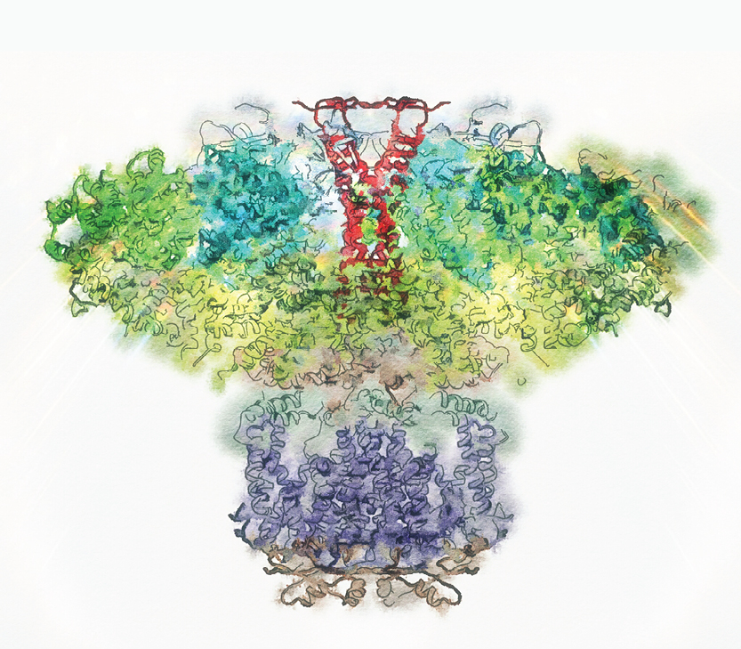
Cryo-EM enables the study of the three-dimensional shapes of biomolecules in their native aqueous environment, and ultimately, to study them in membranes and in their cellular context.
“Whatever we see happening in the natural world is due to the dynamics of life,” said Irina Serysheva, PhD, professor of biochemistry and molecular biology and director, Structural Biology Imaging Center. “Cryo-EM provides snapshots of dynamic proteins, nucleic acids, and complex biological assemblies involved in human health and diseases.”
Structural biologists have employed the Cryo-EM technology at McGovern Medical School since 2004 to understand protein nanomachines related to Alzheimer’s disease, cancer, vision, neurodegeneration, virus infection, cell-cycle regulators, circadian clocks, immune response regulators, and bacterial secretion systems, among other areas.
“The positive impact of our institutional investment in Cryo-EM is evident from the well-funded research projects and high-profile papers that have resulted,” said Rodney Kellems, PhD, professor and chair of the Department of Biochemistry and Molecular Biology. “Because this technology is highly automated, it can support the structural biology research of numerous investigators at McGovern Medical School as well as colleagues from neighboring institutions, thereby promoting collaboration and bringing recognition.”
Led by Serysheva, the Cryo-EM Core Facility houses the most robust Cryo-EM technology in the Houston area.
“Due to the foresight of the Cryo-EM research community at UTHealth Houston, and because of the institutional investment in the high-throughput, high-resolution Titan Krios G3 electron cryomicroscope, UTHealth Houston has emerged as a prominent site for single-particle Cryo-EM and for in situ structural biology via electron cryo-tomography,” said Kellems, the Bob and Hazel Casey Chair in Biochemistry.
Using this state-of-the-art instrument, a growing community of structural biologists at UTHealth Houston and the surrounding area have an opportunity to further exploit the enormous potential of Cryo-EM technology. Users of the Cryo-EM Facility include nearly a dozen faculty members from the Departments of Biochemistry and Molecular Biology, Microbiology and Molecular Genetics, Neurobiology and Anatomy, Neurology, and Integrative Biology and Pharmacology. The combined National Institutes of Health funding of these investigators is over $10 million in FY21.
Powered by biological questions and technical developments, the Cryo-EM Facility at UTHealth Houston is continuously expanding and improving in instrumentation and application. In the past five years or so, Cryo-EM has gone through a revolution made possible by improvements in microscope and camera technology.
Serysheva recently led the application for a $2 million High-End Instrumentation Grant from the NIH that will support the purchase of an additional Cryo-EM, offering significant technological improvements and meeting the increasing demand for these innovative instruments.