Human Brain Organoids in Mice: A New Window Into Brain Development and Psychiatric Disease
 What happens when human-derived brain tissue is allowed to develop within a living nervous system? A new study published in Nature offers an important experimental answer—and opens new possibilities for studying the biology of neurodevelopmental and psychiatric disorders.
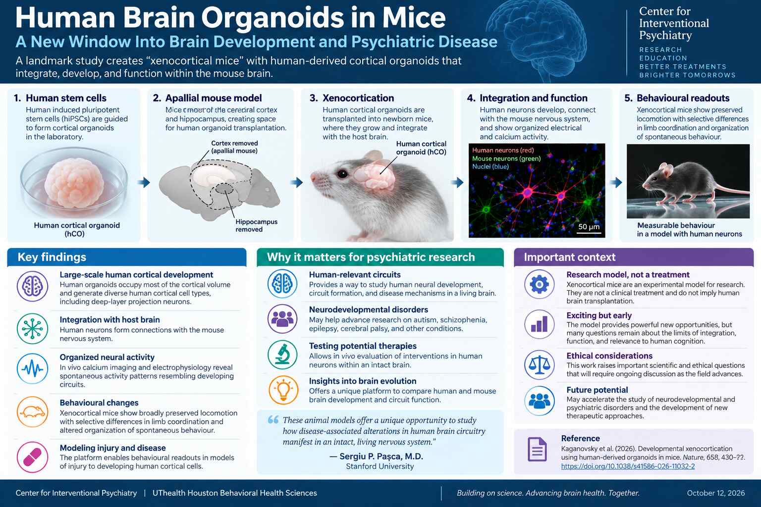
In “Developmental xenocortication using human-derived organoids in mice,” Konstantin Kaganovsky and colleagues, led by Sergiu P. Pașca at Stanford University, developed an approach they call xenocortication. They transplanted human stem-cell-derived cortical organoids into mice genetically engineered to lack most of the neocortex and hippocampus. The resulting human-derived tissue grew, developed specialized cell populations, connected with host circuits, and exhibited coordinated electrical activity.
The central advance is not the creation of a “human brain” inside a mouse. It is a new way to study developing human cortical cells within a living organism, where neural activity can be related to measurable behavior.
Why Existing Brain Organoid Models Have Limitations
Human cortical organoids are three-dimensional structures grown from stem cells that reproduce selected features of developing human brain tissue. They have become powerful tools for studying neuronal differentiation, genetic disease, and developmental biology.
Yet organoids grown in laboratory dishes lack many of the inputs, outputs, and physiological conditions of a living brain. Transplanting organoids into animals can improve maturation, but an intact host cortex limits the physical space available and competes with the human graft for neural connections.
The Stanford group addressed these limitations by creating an immunodeficient mouse model with extensive developmental depletion of cortical and hippocampal tissue, then transplanting cortical organoids into the available space early in life.
What the Researchers Found
The transplanted human cortical organoids survived in 86.2% of 29 assessed mice. By three months after transplantation, the graft accounted for approximately 91.9% of the combined cortical tissue volume in the engrafted animals measured. This is a measure of cortical tissue volume in the experimental mice—not a claim that their entire brains were predominantly human.
Single-nucleus sequencing and spatial analyses identified diverse developing human cortical cell populations. Notably, the grafts included layer 5 extratelencephalic projection neurons, a population difficult to generate in conventional organoid cultures, and a small number of cells with morphology resembling von Economo neurons. The latter should not be equated with fully mature, functionally validated human von Economo neurons.
Tracing studies revealed human-derived projections within the mouse brain and sparse projections extending into the cervical spinal cord. Calcium imaging and electrophysiology showed coordinated, spontaneous activity across the graft, consistent with a developing neural network.
Behavioral results were nuanced. Gross locomotion was broadly preserved, while movement organization and certain cognitive measures differed across control mice, mice lacking much of their cortex, and xenocortical mice. In a Y-maze working-memory task, control and xenocortical mice performed above chance, whereas the non-transplanted apallial mice did not. These findings do not demonstrate that human neurons restored normal mouse cognition.
A Model of Injury to Developing Human Brain Tissue
The investigators then tested whether the platform could register consequences of injury. Following a controlled low-oxygen exposure, human-derived grafts showed a hypoxia-associated cellular response, changes in glial cells, and altered MRI signals. The xenocortical mice also displayed measurable changes in gait and paw support.
This experiment provides an early proof of concept for connecting a biological insult to cellular and behavioral readouts in living animals containing developing human neural tissue. It may be relevant to studying developmental hypoxic injury, but it is not itself a validated model of cerebral palsy or a test of a clinical therapy.
What This Could Mean for Psychiatry
Many psychiatric and neurodevelopmental disorders involve changes in neural development, cellular identity, connectivity, and circuit function. Studying these processes in human-derived cells is important because some features of human cortical development are not adequately reproduced in rodents.
In the longer term, xenocortication could enable investigators to study patient-derived organoids carrying disease-associated genetic variants, observe how these cells interact with circuits in vivo, and test candidate treatments using both cellular and functional measurements. Potential research applications include autism spectrum disorder, schizophrenia, epilepsy, and certain neurodevelopmental syndromes. These are possibilities for future experiments, not diseases shown to be reproduced or treated in this study.
For interventional psychiatry, the broader implication is methodological: meaningful treatment development requires connecting molecular and cellular findings to circuit-level function and, ultimately, behavior. Such models may eventually help generate mechanistic hypotheses relevant to neuromodulation and precision psychiatry. The current study did not test ECT, TMS, ketamine, or other psychiatric interventions.
Scientific and Ethical Boundaries
Important limitations remain. The grafts represented an immature stage of human cortical development, lacked consistent mature cortical layering and full inhibitory-cell representation, and developed within a mouse nervous system that matures on a different timetable. Although the grafts were electrically active and anatomically connected, the investigators could not establish whether graft neurons were necessary or sufficient for specific behaviors.
Human neural tissue in animal hosts also raises questions about animal welfare, altered capacities, and appropriate research oversight. The authors describe institutional and independent ethical review and emphasize the importance of continued engagement with ethicists, patient representatives, and regulatory bodies as these models advance.
It is essential not to confuse this experimental platform with a transplant treatment for patients or to imply that the mice possess human-like cognition or consciousness. Neither conclusion is supported by the study.
The UTHealth Houston Perspective
At the Center for Interventional Psychiatry, advances such as this highlight why the future of psychiatric therapeutics will depend on understanding how cellular mechanisms give rise to functional neural circuits. We increasingly seek to connect biological mechanisms, clinical phenotypes, and treatment response rather than studying each in isolation.
Xenocortication is not yet a clinical technology. Its value lies in potentially enabling more informative preclinical experiments with human-derived neural cells—an important step toward mechanistically grounded approaches to severe and treatment-resistant psychiatric disorders.
Looking Ahead
The next questions are clear: Can the human-derived grafts mature further? Can investigators demonstrate causal links between specific graft circuits and behaviors? Can patient-derived models reproduce meaningful disease features? And can interventions reverse those features in ways that translate to patients?
The answers will require substantial further research. For now, this Nature publication provides a compelling demonstration that developing human cortical tissue can be studied at cellular, network, and behavioral levels within a living experimental system.
Reference
Kaganovsky K, Kelley KW, Gschwind T, et al. Developmental xenocortication using human-derived organoids in mice. Nature. 2026;658:430–441. Published online September 16, 2026. https://doi.org/10.1038/s41586-026-11032-2
Additional context: Brain & Behavior Research Foundation. “Important Advance in Brain Organoid Technology Provides Platform to Study Pathology and Test Therapies…” October 8, 2026.
Contact
Center for Interventional Psychiatry
UTHealth Houston Behavioral Health Sciences
Request for Second Opinion: https://Go.uth.edu/CIPIntake
Phone: (713) 486-2621
Fax: (713) 500-2728
Email: [email protected]
Website: https://go.uth.edu/CIP
Disclaimer
This commentary is for educational purposes and does not constitute medical advice. The reported work is preclinical research and does not establish a treatment for psychiatric or neurological disorders.
AI-assisted drafting disclosure: Artificial intelligence was used as a scientific writing support tool. The final text has been reviewed and approved by João L. de Quevedo, MD, PhD.