Microscopy Core

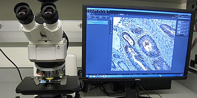
The IMM Microscopy Core provides assistance in confocal microscopy, wide-field fluorescence microscopy, brightfield microscopy and image analysis. The facility is equipped with a Leica TSC SP5 upright confocal microscope with conventional and resonant scanner, a Nikon Eclipse TE2000E inverted wide-field microscope, a Zeiss Axio Scope brightfield microscope, and a computer workstation running Amira software for post-acquisition analysis of imaging data.

The Microscopy Core will support the research needs of IMM research investigators by providing microscopy technical support, training and consultation.

Leica w2

Leica TCS SP5 Confocal Microscope

The Leica TCS SP5 upright confocal microscope is a point scanning confocal microscope with tandem scanners. The resonant scanner enables high speed imaging at frame rates up to 25 fps at 512 x 512 pixels, and the conventional scanner allows high resolution imaging at frame sizes up to 8192 x 8192 pixels (64 Megapixels).


Key features include:

  • Laser illumination lines: Argon 458/476/488/514nm, DPSS 561nm, HeNe 594nm, and HeNe 633nm.
  • AOTF selection and attenuation of laser lines; capable of Region of Interest (ROI) scan.
  • Spectral detection in individually tunable wavelength bands.
  • Three photomultiplier tubes (PMTs) for fluorescence detection and one for transmitted light (bright field, differential interference contrast (DIC)).
  • Software dye separation function.
  • Plan Apo objectives optimized for confocal scanning: 10x (NA 0.4) and 20x (NA 0.7) dry, 40x (NA 1.25) and 63x (NA 1.4) oil, and 63x (NA 1.2) water. The 63x water objective has a correction collar.

Notes on sample preparation for the confocal microscope:

  • Our confocal microscope does not have a source to excite DAPI or Hoechst. Nucleic acid stains to be used with this microscope include but are not limited to: SYTOX Green, SYTOX Orange, CyTRAK Orange, Draq5, TO-PRO-3.
  • Samples need to be mounted in appropriate medium and fluorescence protected by anti-fade reagents.
  • Samples need to be covered with 0.17mm thick coverslips. The coverslip should be sealed securely to the slide/dish holding the sample.
  • To ensure that your sample will work with the confocal microscope, prospective users please discuss your imaging experiment with service center manager before preparing your sample.


Microscopy 1

Nikon Eclipse TE2000E Widefield Fluorescence Microscope

The Nikon Eclipse TE2000E inverted microscope provides for widefield fluorescence and transmitted light imaging. Fluorescence filter sets include DAPI, CFP, FITC, YFP, Texas Red, and Cy5.

Key features include:

  • High speed Prior Scientific excitation and emission filter wheels.
  • A high sensitivity monochromatic Photometrics Cascade 512B EMCCD camera.
  • A high resolution monochromatic Photometrics Coolsnap HQ2 camera.
  • Plan Fluor 4x (NA 0.13) objective.
  • Plan Fluor 10x (NA 0.3) and 20x (NA 0.45) long working distance objectives with phase contrast, for viewing cells through plastic containers. The 20x objective has a correction collar.
  • Plan Apo 40x (NA 0.95) dry and 60x (NA 1.4) oil objectives for viewing samples through coverslip. The 40x objective has a correction collar and a DIC prism.


Imaging Center Brightfield Microscope

Zeiss Axio Scope Brightfield Microscope

The upright Zeiss Axio Scope provides for transmitted light brightfield imaging of histology and immunohistochemistry samples.

Key features include:

  • Axiocam 506 color camera. The 6 megapixel color camera takes images of large sample areas. It offers a dynamic range of 1:2500, making the finest color differences and tiniest details visible in lossless quality. The Peltier cooling guarantees low-noise and reproducible color image quality.
  • Optional 0.63x adaptor further allows the camera to take images of extra-large sample areas.
  • EC Plan-Neofluar 5x (NA 0.16) objective. EC Plan-Neofluar 10x (NA 0.3) objective. EC Plan-Neofluar 20x (NA 0.5) objective. EC Plan-Neofluar 40x (NA 1.3) oil objective. Plan-Apochromat 100x (NA 1.4) oil objective.


Microscopy 2

Image Analysis Workstation with Amira Software

A dedicated computer workstation runs the latest version of Amira software and enables advanced visualization, analysis and presentation of imaging data. Users are welcome to bring images acquired from any microscope (in our facility or elsewhere). No previous experience with the software is required. Assistance is available upon request.

Key features include:

  • Interactive 3D navigation
  • Orthogonal and oblique slicing
  • Volume rendering
  • Surface rendering
  • Deconvolution
  • Segmentation
  • Skeletonization and tracing of neural and vascular networks
  • Image registration
  • Quantifications
  • Co-localization analysis
  • Multi-component analysis
  • Animation tools and movie generation

Acknowledgements in publications demonstrate the value of our core and help us obtain financial and other support. So that we may continue to serve you in the best possible way, investigators who made use of our equipment and services should please acknowledge the IMM Microscopy Core in your publications and notify us of such acknowledgements.


Contacts:

Zhengmei Mao, PhD, Manager
Phone: 713.500.3389 | Email: Zhengmei.Mao@uth.tmc.edu

UTHEALTH
The Brown Foundation Institute Of Molecular Medicine
1825 Pressler Street SRB 616
Houston, Texas 77030