Diagnosing Placenta Accreta
Edgar Hernandez-Andrade, MD, PhD, professor in the Division of Maternal-Fetal Medicine (MFM) at McGovern Medical School at UTHealth Houston, and Eleazar Soto Torres, MD, assistant professor in the division, have imaged and studied placentas together for 14 years, first at Wayne State University and the Perinatology Research Branch of the National Institute of Child Health and Human Development (NICHD) in Detroit, and since 2020 at UTHealth Houston. The two experts in maternal and fetal imaging are intrigued by the function of the placenta itself and have conducted research to characterize it.

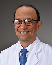
Edgar Hernandez-Andrade, MD, PhD
“The advanced ultrasound technology we use allows us to communicate with the fetus and placenta and better understand what happens before birth,” says Hernandez, who is renowned in his field and the editor-in-chief of Fetal Diagnosis & Therapy. “Without all these advances in ultrasound, obstetrics would be virtually the same as it was 100 years ago.”
Working together, Hernandez and Soto evaluate patients at risk for placenta accreta – those who have been diagnosed with placenta previa and/or have had multiple C-sections or uterine surgeries – to determine the level of risk for the life-threatening condition. They scan patients using various ultrasound techniques to evaluate placental characteristics and vascularity, the relationship between placenta and uterine wall and placenta and pelvic organs, and to quantify blood flow. They evaluate ultrasound images in detail, discuss cases extensively, and reach a diagnosis. Complementary ultrasound techniques – color Doppler, pulsed Doppler, and 3D sonography – allow them to better visualize the uterus, placenta, and surrounding organs in minute detail to determine the difference between a normal placenta and placenta accreta.

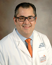
Eleazar E. Soto Torres, MD
Three-dimensional ultrasound provides a view of the placenta from different perspectives, delivering a more accurate diagnosis. The two specialists use MRI as a complementary diagnostic technique, ordering it when they suspect severe cases with bladder and bowel involvement. After diagnosis, they follow each patient with frequent ultrasounds during pregnancy and after delivery if the placenta is left in place, until it resolves.
“All academic centers have similar technology. What sets us apart are the years of experience we have working together and our interest in the placenta itself, which often is overlooked,” Soto says. “Placentas intrigue us. As a result, we see things in a different way and have started thinking outside the box.”
Years of clinical experience and research led Hernandez and Soto to investigate the placenta as a functional unit. “We’re applying these more advanced techniques to show the ways in which the functional unit is abnormal in patients with placenta accreta,” Hernandez says. “As technology advances, we have been able to get a better view of the organs close to the placenta. For instance, we can now evaluate the bladder wall in great detail. Ultrasound systems continue to improve, and we can detect slow blood movements and determine whether vessels are in the bladder or in the placenta.”
Hernandez remains involved with NICHD’s Perinatology Research Branch through inter-institutional collaborations and lectures on perinatology. At UTHealth Houston, he and Soto are investigating the association between placenta accreta and other complications of pregnancy such as preeclampsia and abnormal fetal growth. They are also conducting research that evaluates changes in vasculature and blood flow after delivery in cases when the placenta is left to reabsorb.