UTHealth Ultrasound
ABOUT:
Welcome to the Center For Advanced Ultrasound Imaging Translational Research and Innovations (UTHealthSonix).
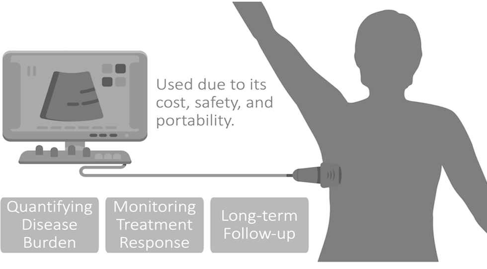
Ultrasound is an inexpensive, portable, and ubiquitous technology that is used to monitor different diseases. Due to its portability, ultrasound equipment can be taken right into the patient room, and modern ultrasound scanners can be used as point-of-care devices connected to small portable systems. Particularly in remote areas with limited access to X-ray and CT, clinicians can sometimes only perform physical examination. Therefore, ultrasound has the potential to be a useful diagnostic tool in such conditions.
The Center for Advanced Ultrasound Imaging Translational Research and Innovations (UTHealthSonix) aims to facilitate and advance the cutting-edge pre-clinical and clinical translational research, and engineering innovation in the field of medical ultrasound for both pre-clinical and clinical applications. The center develops diagnostic and therapeutic technologies for preclinical and clinical applications. Ultimately, the center aims to transfer our technologies to clinical studies and commercialize them. The Center promotes collaborations between various research programs (pediatric and adults) including translational abdominal imaging, neuroimaging, neuroscience, bioinformatics and computational medicine, to name only few.
The director of the ultrasound center, Dr. Alireza Akhbardeh, and core members, Dr. Susan John chair, and Dr. Michael A. Jacobs, vice Chair of Research at the Diagnostic and Interventional Imaging Department, have long track records of publishing and obtaining federal grant funding in the field of ultrasound imaging, other imaging modalities and following applications, to name only a few:
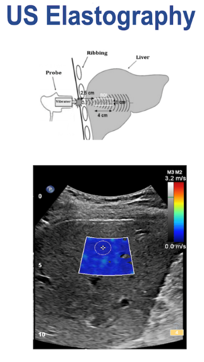
- Liver Fibrosis Staging Using Shear-Wave Elastography and advanced Artificial Intelligence Techniques.
- Liver Fat Quantification Using Shear-Wave Elastography and advanced Artificial Intelligence Techniques.
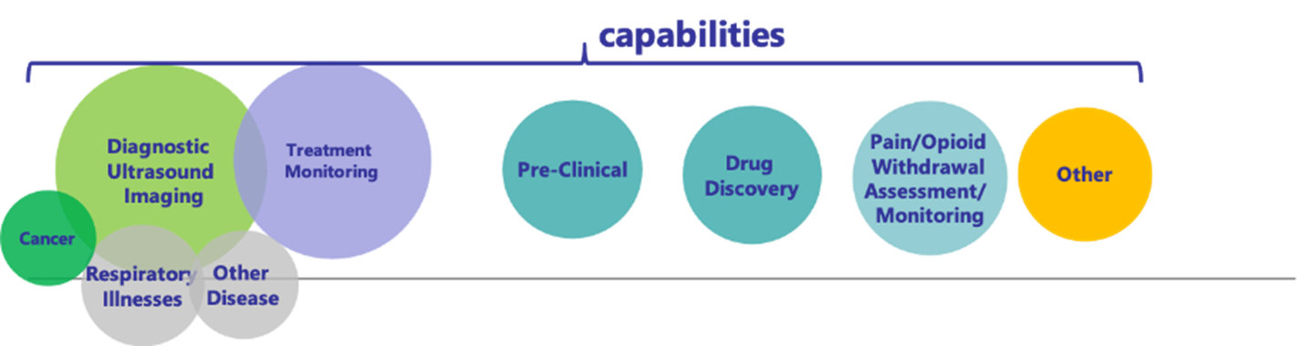
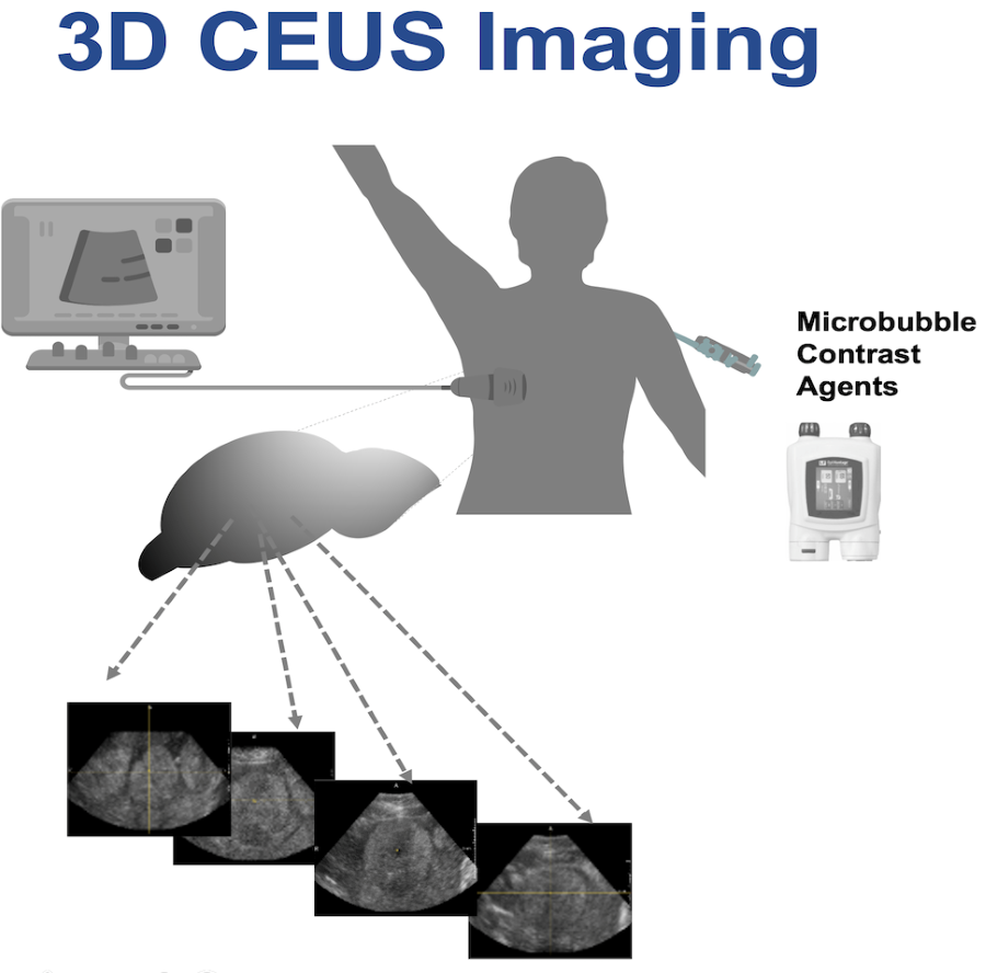
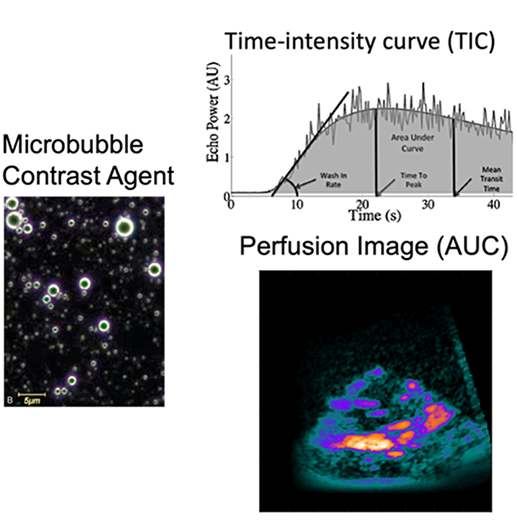
- Early Cancer Diagnosis using 2D and 3D Contrast Ultrasound Imaging (CEUS).
- Automated Lung Ultrasound Quantification for Treatment Effectiveness Monitoring of Patients with Respiratory Illness.
- Quantify bone regeneration using ultrasound-based imaging markers.
- Investigate the use of High Intensity Ultrasound Sound (HIFU) in different diseases.
UTHealthSonix facilitates collaboration between faculty from diverse research domains who are committed to developing this integrative research program to obtain additional extramural funding support. The center core members are actively involved in graduate as well as resident education, and in multiple committees in the university, nationally and internationally. The ultrasound center emphasizes an extensive research experience within a state-of-the-art research environment and aims to provide services, research and development, innovations, training and education for the next generation of translational researchers.
LOCATION:
The Center is located in the at Diagnostic and Interventional Imaging Department, McGovern Medical School in the Medical School Extension (MSE) Building.
CONTACT US:
Please send general questions to UTHealthSonix@uth.tmc.edu or the director of the ultrasound center, Dr. Alireza Akhbardeh. We welcome STEM and medical students interested in ultrasound research.
MISSION AND VISION
The main mission of the Center for Advanced Ultrasound Imaging Translational Research and Innovations (UTHealthSonix) at the McGovern Medical School at the University of Texas Health Science Center at Houston (UTHealth) are:
- Provide Ultrasound Imaging service to all faculty at UTHealth for both pre-clinical and clinical applications.
- Develop diagnostic and therapeutic technologies for preclinical and clinical applications.
- Further and promote Translational research and engineering innovation in the field of medical ultrasound.
- We aim to transfer our technologies to clinical studies and commercialize them.
Additionally, UTHealthSonix center aims to be a state-of-the-art research center in Translational Research and Innovations in the field of ultrasound imaging. The interdisciplinary environment at the Center integrates both basic and translational research programs to provide an outstanding atmosphere for scientific exploration in the field of ultrasound imaging. The Center also promotes services, research and development, innovations, training and education to clinicians and researchers to enable them to approach research problems in ultrasound imaging using integrated methodologies as well as develop critical thinking skills. The Center training opportunities focus on essential competencies in translational ultrasound imaging such as key components of translating from preclinical studies to human clinical trials, understanding the significance of scientific rigor, and acknowledging the importance of biomarkers in clinical ultrasound imaging. The Center brings together nationally recognized and highly productive pre-clinical and clinical researchers of different programs who investigate different diseases burdens and mechanisms at the level of patient care as well as in the areas of basic and translational research in ultrasound imaging. The Center leverages its partnerships with other centers in the University such as SBMI to establish collaborative projects for extramural funding. Research areas of focus include:
- Monitoring acute and chronic diseases (diagnosis and treatment monitoring)
- Therapeutic Applications
- Drug Discovery
- Drug Abuse Objective Assessment
- Objective Opioid withdrawal and Effectiveness Monitoring
- Open-loop and Close loop control of medical dose titration and neuro-stimulation for pain management and opioid withdrawal
GOALS
- To conduct and promote translational ultrasound imaging research that yields new insights in the understanding of the neurobiology of psychiatric disorders and into novel treatment approaches.
- To foster collaborations between pre-clinical, translational and clinical research investigators in the field of ultrasound imaging to develop and invent cutting-edge technologies to quantify disease burden, monitor treatment response and long-term follow-up using ultrasound imaging.
- To prepare the next generation of innovative and creative translational researchers, through education, training, and career development, to gain the breadth of knowledge necessary to apply novel ideas and methodologies in tackling different diseases and disorders.
RESEARCH AT UTHEALTHSONIX:
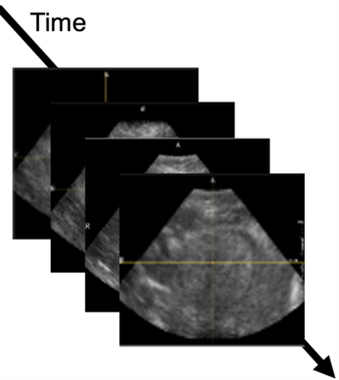
The Center focuses on ultrasound imaging of different organs and biological tissues (pre-clinical and clinical) using the following modes and techniques:
- B-Mode, M-Mode, Doppler imaging.
- Ultrasound Elastography including Shear wave Imaging.
- 2D and 3D Contrast Ultrasound Imaging (CEUS).
- Quantitative Ultrasound Spectroscopic Imaging (QUS) for Characterization of Different Diseases.
UTHealthSonix center houses research and development projects, science communication initiatives, and professional development. The Center has the following Key R&D Applications and Services:
- Conduct sponsored Projects in the following, but not limited to, applications:
- Monitoring acute and chronic diseases (diagnosis and treatment monitoring)
- Therapeutic Applications
- Drug Discovery
- Drug Abuse Objective Assessment
- Objective Opioid withdrawal and Effectiveness Monitoring
- Open-loop and Close loop control of medical dose titration and neuro-stimulation for pain management and opioid withdrawal
- AI- enabled imaging and solutions: One of the key components of this center is the development of advanced Machine Learning and AI to use medical images, clinical and biomedical data for “diagnostics”, “monitoring acute and chronic diseases “, “therapeutic”, and other applications.
- Promote collaborative and interactive research between basic, translational and clinical research investigators to facilitate the research and development of novel application and technologies.
- Promote engagement of research fellows and students who are interested in conducting research activities in the field of ultrasound imaging research and development. In addition, the Center promotes education and training opportunities for residents, fellows and students by organizing bi-weekly invited talks(seminars) and annual workshops on related topics by inviting internationally recognized KOLs (key opinion leaders) from academia and industry.
SELECTED CURRENT AND PREVIOUS PROJECTS:
- Liver Fibrosis Staging Using Shear-Wave Elastography and advance Artificial Intelligence Techniques.
- Liver Fat Quantification Using Shear-Wave Elastography and advance Artificial Intelligence Techniques.
- Early Cancer Diagnosis using 2D and 3D Contrast Ultrasound Imaging (CEUS).
- Automated Lung Ultrasound Quantification for Treatment Effectiveness Monitoring of Patients with Respiratory Illness.
- Quantify bone regeneration using ultrasound-based imaging markers.
- Investigate the use of High Intensity Ultrasound Sound (HIFU) in different diseases.
SELECTED PUBLICATIONS:
- Sagreiya, H.; Jacobs, M.A.; Akhbardeh, A. Automated Lung Ultrasound Pulmonary Disease Quantification Using an Unsupervised Machine Learning Technique for COVID-19. Diagnostics 2023, 13, 2692.
- Akhbardeh, A., Sagreiya, H., Durot, I., and Rubin, D.L. (2021). “Machine Learning for Automated Hepatic Fat Quantification.” TechConnect Briefs 2021, pp105-108.
- El Kaffas, A. Hoogi, J. Zhou, I. Durot, H. Wang, J. Rosenberg, A. Tseng, H. Sagreiya, A. Akhbardeh, D.L. Rubin, A. Kamaya, D. Hristov and J.K. Willmann, Spatial Characterization of Tumor Perfusion Properties from 3D DCE-US Perfusion Maps are Early Predictors of Cancer Treatment Response, Sci Rep 10, 6996, 2020.
- Durot, A. Akhbardeh, H. Sagreiya, A. M. Loening, D.L. Rubin, “A New Multimodel Machine Learning Framework to Improve Hepatic Fibrosis Grading Using Ultrasound Elastography Systems from Different Vendors.” Ultrasound in medicine & biology vol. 46,1 (2020): 26-33.
- Sagreiya, A. Akhbardeh, D. Li, R. Sigrist, B. Chung, G. Sonn, Lu. Tian, J.K. Willmann, “Point Shear Wave Elastography Using Machine Learning to Differentiate Renal Cell Carcinoma and Angiomyolipoma,” Ultrasound Med Biol. 2019 Aug;45(8):1944-1954.
- Akhbardeh, A. El Kaffas, H. Sagreiya, J.K. Willmann, D.L. Rubin, “A multi-model framework to estimate perfusion parameters using contrast-enhanced ultrasound imaging,” Med Phys. 2019 Feb;46(2):590-600.
- Durot, A. Akhbardeh, J. Rosenberg, J. K. Willmann, “Point Shear Wave Elastography for Grading Liver Fibrosis: Can the Number of Measurements be Reduced? ,” Ultrasound Med Biol. 2018 Dec;44(12):2569-2577.
- Jacobs MA, Herskovits EH, Kim HS. Uterine Fibroids: Diffusion-weighted MR Imaging for Monitoring Therapy with Focused Ultrasound Surgery–Preliminary Study. Radiology. 2005;236:196–203.
- Kim HS, Baik JH, Pham LD, Jacobs MA. MR-guided high-intensity focused ultrasound treatment for symptomatic uterine leiomyomata: long-term outcomes. Acad Radiol. 2011 Aug;18(8):970-976.