2026 IMS-iRS Congress Digital Poster Abstracts
Thank you to everyone who submitted an abstract for the 2026 IMS-iRS Congress. Please see the abstracts selected to be displayed during the congress as digital posters below, listed by submitter’s last name.
Abiko, Masaru
Skull Base Vascular Rescue Surgery After Failed Endovascular Treatment: Lessons from Two Complex Posterior Circulation Cases
Masaru Abiko, Koki Ikeda, Ryosuke Dowaki
JA General Hospital
Introduction/Background
Although endovascular therapy has become the primary treatment for posterior circulation aneurysms, complex recurrent lesions occasionally require microsurgical rescue procedures utilizing skull base approaches and cerebral revascularization.
Objectives
To demonstrate the contemporary role of skull base vascular surgery as a rescue strategy after failed endovascular treatment.
Methods/Description
Two patients with recurrent posterior circulation aneurysms following endovascular treatment were reviewed. Case 1 was a ruptured basilar artery fenestration aneurysm treated with coil embolization that subsequently recurred. Direct clipping was performed through a left combined petrosal approach because of a high-riding right jugular bulb. Case 2 was a ruptured vertebral artery dissecting aneurysm initially treated with stent-assisted coiling. Early recurrence developed despite treatment, requiring a V3–radial artery graft–P2 bypass followed by parent artery occlusion.
Results/Comments
In Case 1, complete obliteration of the aneurysm was achieved while preserving perforating arteries, without new neurological deficits. In Case 2, the bypass remained patent and durable exclusion of the aneurysm was obtained. Both patients achieved favorable outcomes. These cases demonstrate scenarios in which definitive treatment could not be achieved by endovascular techniques alone and required advanced skull base exposure and microsurgical reconstruction.
Conclusion
Skull base vascular surgery remains an indispensable rescue option for selected recurrent posterior circulation aneurysms in the endovascular era. Mastery of skull base approaches and bypass techniques continues to be essential for achieving durable cure in complex cases.
Akhtar, Muhammad S.
Who Actually Benefits from Immersive Anatomy? 3D Reconstruction of Routine Imaging and Skull Base Structure Identification Across a Competence Gradient
Muhammad Shaheer Akhtar, Kevin O’Neill
Imperial College NHS Trust London
Introduction
Immersive 3D improves anatomy learning overall (meta-analytic SMD 0.58), but that evidence is mostly general anatomy, and skull base comparisons are qualitative or from cadaveric photogrammetry—which presupposes a dissection laboratory. Cognitive-load theory predicts benefit should narrow as expertise rises; this is untested for skull base anatomy across a training gradient.
Objectives
To measure whether immersive 3D reconstruction of routine imaging improves identification of skull base neuroanatomy, and whether that benefit varies with training level.
Methods/Description
35 participants across three strata—medical students (n=15), junior doctors (n=10), neurosurgical trainees (n=10)—identified skull base structures and corridors (cranial nerves, Meckel’s cave, optic canal, cavernous sinus, internal carotid artery, petrous apex) on conventional 2D CT/MRI and in a purpose-built extended-reality headset showing the identical studies. The same platform generated every model automatically at native intensity, without manual segmentation or cadaveric material. Presentation order was counterbalanced. Primary outcome was identification accuracy; confidence was rated per structure.
Results/Comments
Accuracy improved from 62% to 78% with 3D (p<0.05). Benefit was greatest among medical students (48% vs 72%, p<0.05), intermediate among junior doctors, and smallest among neurosurgical trainees (78% vs 84%, p=0.09). Confidence rose across all strata, including where accuracy did not—a miscalibration relevant to immersive assessment.
Conclusion
Immersive reconstruction of routine imaging improves skull base structure identification, and the benefit is unevenly distributed across competence—with curricular implications for timing. Because one platform builds models from routine scans, not cadavers, this route is available to centres without a dissection laboratory.
Alawieh, Ali
Data-Driven Detection of Neurosurgical Research Eras from 1946 to 2025
Ali Alawieh, Jonathan Grossberg, Eva Wu, Prateek Konakalla, Daniel Barrow, Adam Arthur, Paul Park, Nickalus Khan
Semmes Murphey Clinic
Introduction/Background
Neurosurgery is commonly described through technology-defined eras, but whether those eras can be recovered directly from the publication record has not been established.
Objectives
To reconstruct neurosurgery’s publication history from 1946-2025 and determine whether statistically detected era boundaries align with major clinical and technological milestones.
Methods/Description
Published records with neurosurgery-affiliated authorship were retrieved and expanded to all works by 317,293 seed authors, yielding 18.6 million unique works. Titles and indexed abstracts were classified into 29 neurosurgical subspecialty labels using a curated keyword classifier validated against OpenAlex primary-topic assignments and expert review. Era boundaries were inferred from annual multivariate subspecialty-share matrices using Pruned Exact Linear Time change-point detection with radial basis function cost. Robustness was assessed by penalty sensitivity and moving-block bootstrap.
Results/Comments
PELT identified five boundaries–1964, 1984, 1999, 2019, and 2024–defining six publication eras: classical-open surgical, stereotactic-expansion, MRI era, endovascular/stereotactic-radiosurgery, thrombectomy/molecular-neuro-oncology, and emerging robotics/AI. The 1964 boundary persisted across six of seven penalty settings and had a block-bootstrap 95% interval of 1959-1969. Boundaries in 1984 and 1999 had bootstrap intervals of 1979-1989 and 1994-2004, while 2019 and 2024 were recovered in every replicate. Subspecialty diversity rose from 2.58 bits in 1947 to 3.22 bits in 2017 before declining, and a random-forest model classified eras with 97.1% leave-one-out accuracy.
Conclusion
Neurosurgical eras are statistically recoverable from global publication patterns without pre-specifying milestones, providing a quantitative framework for specialty history and future technology-diffusion benchmarking.
Alsamari, Ali
Benchmarking Meningioma Resection Outcomes by Complex Spine-Trained Neurosurgeons in a Community Hospital
Ali Alsamari, Abdul-Haseeb Naeem, Al-Retage Al-Bader, Amrit Kaur Grewal
Windsor Regional Hospital
Background
Community hospitals often encounter challenges delivering specialized neurosurgical care. The complexity of meningioma resections can lead to suboptimal outcomes due to procedures falling outside a surgeon’s fellowship training. Objective: This study evaluates clinical outcomes following meningioma resection at a community hospital, benchmarking these results against national data.
Methods
A retrospective chart review of 78 patients who underwent meningioma resection at Windsor Regional Hospital (2021–2024). Data analyzed included demographics, tumor characteristics (grade/location), extent of resection (GTR/STR), complications, adjuvant therapies, and pre- and post-operative neurological status.
Results
The cohort (mean age 61, median 65) comprised 63 Grade 1 (M1) and 15 Grade 2 (M2) meningiomas. Locations were predominantly frontal (64.48%). Gross total resection was achieved in 46/63 M1 and 12/15 M2 cases. Postoperative neurological status improved in 42% of patients, remained stable in 38%, and declined in 20%. Complication rates were low, with infections and delirium (7.69%) and hemiparesis (6.41%) being the most frequent. Adjuvant therapy was limited, with 3 patients receiving radiation and 1 chemotherapy. The observed one- and two-year survival rates were 95.23% and 92.3%, respectively. When compared with national data benchmarks of 92.8% and 90.7%, these figures yield relative one- and two-year survival rates of 1.04 and 1.02, respectively.
Conclusion
Our findings demonstrate that outcomes following meningioma resection in a community setting are consistent with national profiles, suggesting that optimal meningioma outcomes can be achieved in community hospitals, performed by complex spine-trained neurosurgeons.
Araújo, Matheus Assis
Beyond the Classical Circle of Willis: Cadaveric Mapping of Cerebral Arterial Variations in a Brazilian Population
Matheus de Assis Araujo, Elizangela Ferraz Pazinato, Any Kelly Gomes de Lima, Marcio Massao Kawano
Indiana University
Introduction/Background
Cerebral arterial variants alter collateral flow and challenge cerebrovascular procedures. Brazilian cadaveric data remain scarce.
Objectives
To map arterial-circle and vertebrobasilar variations in complementary fresh-cadaver series.
Methods/Description
At a Brazilian forensic service, 34 fresh brains underwent arterial-circle dissection and a 30-brain series underwent vertebral, basilar, anterior inferior cerebellar (AICA), and superior cerebellar (SCA) assessment. Caliber, course, continuity, branching, hypoplasia, duplication, dominance, and basilar curvature were recorded.
Results/Comments
Only 2/34 arterial circles (5.9%) displayed the classical configuration; 32 (94.1%) contained at least one variant. Duplication occurred in 22 specimens, comprising 28 duplicated vessels; the anterior communicating artery was most frequently duplicated (10/34; 29.4%). Seventeen specimens contained 20 absent vessels, 18 involving posterior communicating arteries. Hypoplasia most often affected anterior cerebral arteries. Rare patterns included anterior communicating artery fenestration, anomalously originating and intertwined anterior cerebral arteries, atypical posterior communicating artery courses, and complex bifurcation/trifurcation. In the posterior series, 9/30 specimens (30.0%) had at least one variant. Among 25 evaluable vertebral pairs, dominance was right-sided in 11, left-sided in 10, and absent in 4; basilar curvature generally deviated contralaterally. AICA variants comprised four absences, two duplications, and one early bifurcation; SCA variants comprised one duplication and one early bifurcation.
Conclusion
Cerebral angioarchitecture was highly variable, particularly in communicating and vertebrobasilar vessels. Meticulous angiographic review is essential before aneurysm, skull-base, posterior-fossa, and endovascular procedures.
Araújo, Matheus Assis
Burr-Hole-Free Osteoplastic Supraorbital Keyhole Clipping of Anterior Circulation Aneurysms: A Two-Center Brazilian Experience
Matheus de Assis Araújo, Felippe Antunes, Carlos Eduardo Ferrarez, Luiz Otávio Sales Pimenta
Indiana University
Introduction/Background
Evidence supporting supraorbital keyhole aneurysm clipping in public hospitals remains limited.
Objectives
To assess angiographic, surgical, functional, and cosmetic outcomes of burr-hole-free osteoplastic supraorbital clipping at two Brazilian public hospitals.
Methods/Description
Consecutive patients undergoing this technique between January 2017 and December 2025 were retrospectively reviewed. Endpoints included postoperative aneurysm occlusion, modified Rankin Scale (mRS) 0–2 at 6 months, conversion, complications, mortality, recurrence, and cosmesis.
Results/Comments
Sixty-four patients with 72 anterior circulation aneurysms were included; 45 (70.3%) presented after rupture. Aneurysms arose from the anterior communicating artery in 50 (69.4%) and posterior communicating artery in 14 (19.4%); mean diameter was 6.8±2.9 mm. Mean operative time was 172±39 minutes and blood loss 190±110 mL. Temporary clipping was used in 14 patients (21.9%), intraoperative rupture occurred in 4 (6.3%), and no case required conversion. Complete occlusion was achieved in 69/72 aneurysms (95.8%). Procedure-related infarction and permanent neurological deficit each occurred in 2 patients (3.1%); one hematoma required evacuation (1.6%). No cerebrospinal fluid leak, optic nerve injury, or approach-related death occurred. At 6 months, 58 patients (90.6%) had mRS 0–2. Overall mortality was 3.1%, exclusively after severe ruptured aneurysms. Cosmetic assessment was available in 62 survivors, all reporting good/excellent results. One recurrence (1.4%) occurred during 30 months’ mean follow-up.
Conclusion
In selected anterior circulation aneurysms, this two-center approach achieved high occlusion, favorable functional recovery, and excellent cosmesis without conversion. Functional outcome appeared primarily influenced by initial hemorrhage severity.
Araújo, Matheus Assis
Intracranial Meningiomas in Regional Brazilian Hospitals: Surgical Profile, Extent of Resection, and Early Outcomes
Matheus de Assis Araújo, Felippe Antunes, Carlos Eduardo Ferrarez, Luiz Otavio Sales Pimenta
Indiana University
Introduction/Background
Data from regional Brazilian neurosurgical centers on meningioma surgery and outcomes remain limited.
Objectives
To characterize the epidemiological, anatomical, histopathological, surgical, and follow-up profile of intracranial meningiomas treated at two regional hospitals.
Methods/Description
We retrospectively reviewed 63 patients with 68 surgically treated intracranial meningiomas in Minas Gerais, Brazil, from January 2021 to February 2026. Demographics, location, WHO and Simpson grades, complications, neurological deficits, reoperations, and recurrence were analyzed. Fifty-four patients had follow-up through December 2025; 2026 cases remain under surveillance.
Results/Comments
Forty-two patients were women (66.7%); mean age was 48 years (range, 29–69). Tumors involved the convexity (22), parasagittal region (12), olfactory groove (11), sphenoid wing (8), tuberculum sellae (7), petroclival region (5), and tentorium/posterior fossa (3). WHO grades 1, 2, and 3 comprised 63 (92.6%), 3 (4.4%), and 2 (2.9%) tumors. Simpson grades 1 and 2 were achieved in 19 (27.9%) and 31 (45.6%); thus, 50/68 (73.5%) underwent Simpson 1–2 resection and 18 (26.5%) Simpson 3–4 resection. Seven cerebrospinal fluid leaks and six infections occurred. Ten patients (15.9%) developed new neurological deficits; eight recovered completely within a mean of 60 days, whereas two retained mild, non-disabling upper-limb weakness. Five recurrences/progressions occurred among 51 evaluable cases (9.8%); two were reoperated and three await reintervention. No surgery-related deaths occurred.
Conclusion
Regional centers achieved predominantly WHO grade 1 pathology and favorable neurological recovery. Continued surveillance is warranted, particularly after Simpson 3–4 resections.
Bcharah, Estefana
Outcomes of Meningiomas Treated by Transbasal Approach Variants: A Scoping Review of the Literature
Estefana Bcharah, Matthew Dufault, Evelyn Turcotte, Alice Giotta Lucifero, Amirhossein Akhavan-Sigari, Landon Gray, Alexandre Bossi Todeschini, Charbel Moussalem, Bernard Bendok
Mayo Clinic
Introduction/Background
Anterior cranial base meningiomas, including olfactory groove, planum sphenoidale, and tuberculum sellae lesions, often grow on the anterior skull base and invade the orbit and paranasal sinuses before diagnosis. The transbasal approach, a midline extradural corridor, reaches the anterior fossa floor and parasellar region with minimal frontal lobe retraction, though its optimal configuration remains debated.
Objectives
To characterize published experience with the transbasal approach for meningioma and compare its standard and extended variants.
Methods/Description
A search of four databases (PubMed, Web of Science, Cochrane, Embase) using “transbasal,” “approach,” “meningioma” and equivalents identified studies of transbasally treated patients. Approaches classified as standard transbasal (suprasinus or transsinus) or extended transbasal with orbital bar or rim removal were included.
Results/Comments
Twenty-five studies yielded 372 patients. Mean age was 53.2 years and 63.5% were female. Of those with a reported subtype, 71.8% were olfactory groove and 26.6% were tuberculum sellae meningiomas.
The most common presentation was cognitive change (38.0%), followed by headache (35.4%), visual impairment (32.0%), and olfactory dysfunction (29.1%). The standard approach addressed midline tumors without paranasal extension, whereas the extended approach addressed larger tumors with paranasal sinus, orbital, and extracranial bony invasion.
Gross total resection was achieved in 91.9% of cases, and visual function improved in 64.8% of patients with preoperative deficits; where fully reported, 56.5% improved, 34.8% remained stable, and 8.7% worsened.
Conclusion
The transbasal approach achieves high resection rates for large midline meningiomas. Selecting standard versus extended variants should be individualized to tumor size, vascular relationships, and sinonasal extension.
Becker, Hannes
Petroclival and Sphenopetroclival Meningiomas: Surgical Management, functional Outcome, and long-Term Follow-up.
Hannes Becker, Christina Fodi, Hanna Goett, Jonas Tellermann, Nathalie Arzer, Peter Paßlack, Jens Schittenhelm, Jürgen Honegger, Felix Behling, Marcos Tatagiba
Department of Neurosurgery, University Hospital Tuebingen, Eberhard Karls University Tuebingen
Introduction/Background
Petroclival and sphenopetroclival meningiomas are rare skull base tumors associated with considerable surgical complexity and morbidity because of their proximity to critical cranial nerves and neurovascular structures. Optimal treatment strategies remain a matter of debate.
Objectives
To evaluate clinical characteristics, surgical management, neurological outcomes in patients with petroclival and sphenopetroclival meningiomas.
Methods/Description
We retrospectively analyzed patients treated for petroclival and sphenopetroclival meningiomas at a tertiary skull base center between 2003 and 2022. Clinical presentation, surgical strategy, extent of resection, histopathology, and functional outcomes were assessed.
Results/ Comments
Among 2,603 meningioma cases, 78 tumors met the inclusion criteria, comprising 58 petroclival and 20 sphenopetroclival meningiomas. Patients were predominantly female with a mean age of 56.3 years. Facial nerve dysfunction and dysphagia were the most common preoperative symptoms. Most tumors were resected via a retrosigmoid approach, while selected sphenopetroclival tumors required a two-stage procedure. Gross-total resection was achieved in 31.1% of petroclival meningiomas, whereas all sphenopetroclival tumors required subtotal resection. WHO grade 2 histology was more frequent in sphenopetroclival than petroclival tumors (15.0% vs. 3.4%). Although neurological deficits often worsened transiently postoperatively, long-term follow-up demonstrated improvement in facial nerve function. After a mean follow-up of 52.4 months, recurrence rates were 27.6% for petroclival and 65.0% for sphenopetroclival meningiomas with significantly shorter symptom-free survival in the latter group.
Conclusion
Surgical outcomes were more favorable for petroclival than sphenopetroclival meningiomas, reflecting differences in microsurgical accessibility and tumor biology. These findings highlight the need for individualized treatment strategies tailored to tumor location and characteristics.
Bektasoglu, Pınar Kuru
Microsurgical anatomy of the anterior commissure with special emphasis on the anterior limb: a comparative white matter fiber microdissection and tractography study
Pinar Kuru Bektasoglu, Serdar Rahmanov, Abuzer Gungor, Sima Cebecik Cakir, Zeynep Firat, Fuat Pirincci, Erhan Celikoglu, Wolfgang J. Weninger, Uğur Türe
Istanbul Fatih Sultan Mehmet Education and Research Hospital
Introduction
The anterior commissure (AC) has an anterior and posterior limb. Despite comprehensive information about the posterior limb, there is limited and conflicting information about the anterior limb in the literature.
Objectives
We aimed to demonstrate the anatomical relationships of the AC with neighboring structures using white matter microdissection and magnetic resonance (MR) tractography, with a primary focus on the anterior limb of the AC.
Methods
Twenty cadaveric human brains and three sheep brains were prepared according to Klingler’s method. White matter fiber microdissections were performed. MR tractography was done in 20 specimens.
Results
This study defined the body and anterior and posterior limbs of the AC. The anterior limb leaves the body from the inferior side at the level of the anterior perforated substance and olfactory tract. It then courses anteroinferolaterally, curves towards the orbital gyri, and terminates at the olfactory trigone. The posterior limb of the AC divides into rostral (anterolateral) and caudal (posterolateral) parts. The anterior limb was more prominent in sheep than in human brains.
Conclusion
This study accurately delineates the anatomy and variations of the anterior limb of the AC in human and sheep brains, to the best of our knowledge, for the first time in the literature. The anterior limb of the AC is larger in sheep than in humans. An increased importance of olfaction could be the evolutionary explanation for this difference. The MR tractography results shown are unique for this delicate anatomy.
Keywords
Amygdala; Anterior commissure; Anterior limb; Olfactory tract; Posterior limb.
Brehar, Felix
Lessons from Glioblastoma: A Translational Multi-Omics Framework for High-Grade Meningioma Recurrence
Felix BREHAR, Alexandra Mihaela Patrascan, George Emil Dragos Petrescu, Viorel Mihai Pruna
”Bagdasar-Arseni” Clinical Hospital, ”Carol Davila” University of Medicine and Pharmacy
ABSTRACT
Background
The recurrence of high-grade meningiomas (WHO grades 2 and 3) represents a major challenge for neurosurgeons. Molecular mechanisms involved in glioblastoma progression are documented through numerous genomic, proteomic, and metabolomic studies. Therefore, research protocols investigating glioblastoma recurrence could serve as a model to reveal the molecular mechanisms involved in high-grade meningioma recurrence.
Objective
To propose an intertumoral translational research framework for high-grade meningioma recurrence.
Methods
As a model for high-grade meningioma recurrence research, we propose the research protocol developed for identifying new molecular markers and targets in glioblastoma recurrence within the PerCareGlio project. Tumor samples from initial (iGBM) and recurrent (rGBM) glioblastomas were collected during surgical resection. Additionally, serial peripheral blood samples were collected preoperatively, 5 days postoperatively, and monthly thereafter until documented recurrence. Molecular analyses included miRNA profiling, plasma proteomics and metabolomics using mass spectrometry, followed by multi-omics data integration to identify potential biomarkers associated with tumor recurrence.
Results
The PerCareGlio workflow identified several miRNAs associated with tumor recurrence, while proteometabolomic analyses highlighted ASAH1 (acid ceramidase) and SYNM (synemin) as potential plasma biomarkers for monitoring glioblastoma recurrence, demonstrating the feasibility of longitudinal serum monitoring. The research protocol developed within the PerCareGlio project could be adapted to high-grade meningioma recurrence research. Our findings support the implementation of a similar multi-omics approach to capture the molecular landscape involved in meningioma recurrence.
Conclusion
The PerCareGlio research protocol could serve as a translational research framework for identifying early predictive molecular biomarkers of high-grade meningioma recurrence.
Keywords
High-grade meningioma; glioblastoma; research protocols
Disclosure
This work is supported by the grant COFUND-EP-PERMED-PerCareGlio 89/2025 (UEFISCDI).
Chen, Zhengyuan
Endoscopic Transmaxillary and Transpterygoid Approach for Resection of Mesiotemporal Cavernous Malformation
Zhengyuan Chen, Guoqiang Zhao, Shaojian Lin, Filippos-Savvas Chelmis, Garret Choby, Eric Wang, Carl Snyderman, Georgios Zenonos
University of Pittsburgh Medical Center
Abstract
Background
Mesiotemporal cavernous malformations may present with seizures and hemorrhage, but conventional transcranial approaches often require temporal lobe retraction and risk transgressing functional white matter pathways. This operative video demonstrates an endoscopic transmaxillary–transpterygoid corridor for resection of a mesiotemporal cavernous malformation.
Methods
A 23-year-old man presented with a first-time generalized seizure and normal neurological examination. CT, MRI, susceptibility-weighted imaging, contrast-enhanced sequences, and high-definition fiber tractography demonstrated a left mesiotemporal cavernous malformation with associated hematoma and its relationship to the sagittal stratum. An anterior endoscopic corridor was selected to minimize temporal stem injury and craniotomy-related morbidity.
Results
The patient was positioned supine with stereotactic navigation, neurophysiological monitoring, Doppler ultrasonography, and indocyanine-green endoscopy. After preparation of a contralateral nasoseptal flap and ipsilateral reverse flap, a transpterygoid route was combined with Caldwell–Luc anterior maxillary antrostomy. The infraorbital nerve and V2 were identified, dissected, and medially transposed to expand the transmaxillary working corridor. Navigation-guided drilling of the greater sphenoid wing exposed the temporal pole. Doppler guidance localized the hematoma cavity, which was entered and evacuated using a two-surgeon, four-hand endoscopic technique, followed by multilayer skull base reconstruction. The patient recovered without recurrent seizure, facial sensory disturbance, new neurological deficit, or cerebrospinal fluid leak. The lumbar drain was removed on postoperative day 3, and he was discharged on postoperative day 4.
Conclusion
This case illustrates that an endoscopic transmaxillary–transpterygoid approach can provide a direct, coaxial, cosmetically favorable corridor to selected mesiotemporal cavernous malformations while minimizing brain retraction and temporal stem transgression.
Cirillo, Carlo
Adjuvant radiotherapy versus surgery alone for WHO grade III (anaplastic) meningioma: a systematic review and meta‑analysis of survival outcomes
Carlo Cirillo, Ariana Sofia Vergara Fernandes, Mateo Serrano Pinzón
Federico II
Introduction
Grade III meningiomas are a rare and nefarious tumor. Modern-day guidelines (EANO, NCCN) base recommendations largely on grade II information, leaving significant gaps in the literature. Objectives To quantify the effect of adjuvant radiotherapy against surgery alone on adults with newly diagnosed grade III meningioma
Methods/Description
A systematic search of PubMed, Embase, and Cochrane Library up to May 2026 was done. Records were screened independently by two reviewers, who also assessed risk of bias. Eligible studies were comparative (radiotherapy/surgery alone) and reporting overall survival. A random‑effects DerSimonian‑Laird method was used. τ² was estimated by the method of moments; the studies were weighted by the inverse of the sum of within‑study variance plus τ². Heterogeneity was quantified with the I² statistic and Cochran’s Q test. Pre‑specified subgroups were: the extent of resection (gross‑total vs. subtotal) and study design (registry‑based vs. institutional). Sensitivity analyses excluded extreme estimates and univariate‑only studies.
Results/Comments
Twelve studies (n = 1655) were included in the primary meta-analysis. Adjuvant radiotherapy significantly improved overall survival (pooled HR 0.42, 95 % CI 0.29–0.61, p < 0.0001). There was substantial heterogeneity (I² = 71 %, τ² = 0.225, Q p < 0.001). The benefit remained after excluding extreme values (HR 0.48) and univariate studies (HR 0.45). Subgroup analysis showed consistent benefit across resection strata; registry‑based studies showed a smaller effect than institutional cohorts.
Conclusion
Adjuvant radiotherapy confers a clinically significant survival benefit in WHO grade III meningioma, independent of resection extent, addressing a gap in current guidelines.
Ding, Xing Hua
Management Strategies for Incidentally Discovered Small Meningiomas
XingHua Ding, Mingguang Zhang
HuaShan Hospital Fudan University
Background
With the popularization of neuroimaging techniques and population aging, incidentally discovered small intracranial meningiomas have increasingly become a common issue in neurosurgical clinical practice. The vast majority of such tumors are WHO Grade I and grow slowly, yet some cases still carry a risk of progression. Clinical decision-making requires striking a balance between avoiding overtreatment and preventing delayed intervention. This article systematically reviews the epidemiology, natural history, progression predictors, and current mainstream therapeutic strategies (active surveillance, surgical resection, stereotactic radiosurgery) of incidental small meningiomas.
Aim
To discuss the treatment strategies of small meningioma. Methods: This study analyzed the clinical data of small meningiomas of 56 cases treated by microscope keyhole approach between January 2012 and December 2014, observed the curative effects by outpatient follow-up, and combined with literature to discuss treatment strategies for these diseases.
Results
Among 56 cases of patients, there were 15 male patients and 41 female patients, aged 35 to 80 years old, average age 52±10 years old. 35 cases of tumor located in the cerebral hemisphere convex, 4 cases of para-sagittal sinus, 1 case of cerebral falx, 2 cases of cerebellar hemisphere convex, 3 cases of anterior skull base, 6 cases of sellae-region, 2 cases of middle skull base , and 3 cases of cerebellopontine angle. All tumors adopted the keyhole surgery, among them, cerebral hemisphere convex tumors assisted with enhanced MRI neuronavigation. Except meningiomas in the para-sagittal sinus, anterior skull base, sellae-region, middle skull base, and cerebellopontine angle area achieved Simpson grade Ⅱ resection, the others achieved Simpson grade I resection. The postoperative adverse reaction ratio of 2/56 (3.57%). 42 cases received follow-up, the average follow-up time was10 years. It was confirmed by enhanced MRI that there was no tumor recurrence and by physical examination there was no permanent neurological dysfunction.
Conclusion
The keyhole microneurosurgery was a safe and effective way to treat intracranial small meningioma, supporting our opinions of doing early surgical treatment on small meningioma
Dufault, Matthew
Multimodal Cadaveric Mapping of the External and Internal Carotid Arteries Using CTA, Microsurgical Dissection, and 3D Photogrammetry
Matthew Dufault, Alice Giotta Lucifero, Charbel Moussalem, Alexandre Bossi Todeschini, Amirhossein Akhavan-Sigari, Estefana Bcharah, Landon Gray, Evelyn Turcotte, Bernard Bendok
Mayo Clinic Arizona
Introduction
Understanding of carotid anatomy is essential for cranial neurosurgery, endovascular navigation, and neuroanatomical education. Conventional two-dimensional imaging and photographs incompletely represent the three-dimensional relationships between the external carotid artery (ECA), internal carotid artery (ICA), and cranial structures.
Objectives
To establish a reproducible cadaveric workflow for neurovascular study, ECA and ICA dissection, and to create three-dimensional interactive anatomical models using CTA, latex vascular perfusion, microsurgical exposure, and photogrammetry.
Methods
Two fresh-frozen cadaveric heads were prepared with bilateral common carotid artery, vertebral artery, and internal jugular vein cannulation, followed by injection of iodine-based contrast and CTA. Arteries and veins were then injected with colored latex to preserve vascular architecture. Microsurgical dissections exposed the ECA and major branches, cervical ICA, skull base carotid course, and cavernous/paraclinoid ICA. Sequential 3D photogrammetry captured progressive dissection stages and was integrated with CTA reconstructions to generate detailed three-dimensional anatomical models for anatomical study.
Results
Cadaveric specimens demonstrated complete filling of the anterior and posterior circulations with preservation of carotid anatomy. CTA reconstructions and photogrammetric models allowed visualization of ECA branches, ICA segmental anatomy, and surgically relevant neurovascular relationships. The resulting datasets facilitated correlation between radiographic imaging and microsurgical anatomy and supported development of detailed three-dimensional models.
Conclusion
This multimodal cadaveric workflow enables the creation of detailed 3D anatomical models of the ECA and ICA while providing a reproducible framework for neurovascular anatomical study. Integration of radiographic, microsurgical, and three-dimensional visualization techniques may enhance surgical planning, endovascular education, and translational neurovascular research.
Dufault, Matthew
Three-Dimensional Photogrammetric and Angiographic “Circle of Willis” Atlas of the Anterior Skull Base Approaches
Matthew Dufault, Alice Giotta Lucifero, Charbel Moussalem, Alexandre Bossi Todeschini, Amirhossein Akhavan-Sigari, Estefana Bcharah, Landon Gray, Evelyn Turcotte, Bernard Bendok
Mayo Clinic Arizona
Introduction
Anterior skull base approaches require precise understanding of the anterior cranial fossa, olfactory and optic apparatus and the neurovascular relationships. Conventional operative photographs and two-dimensional imaging incompletely represent the depth, bony anatomy, and stepwise surgical corridors encountered during surgery.
Objectives
To develop a reproducible high-definition three-dimensional photogrammetric and angiographic atlas of anterior skull base approaches, combining cadaveric microsurgical dissection, CTA, and sequential high-definition image acquisition for interactive model generation.
Methods
Fresh formalin-perfused cadaveric heads were prepared by cannulating bilateral common carotid arteries, vertebral arteries, and bilateral internal jugular veins. The vessels were injected with iodine-based contrast and underwent high-resolution CTA. Colored latex vascular perfusion was then performed. The anterior skull base dissections were completed in a stepwise fashion including transbasal, supraorbital, lateral supraorbital, extended lateral supraorbital, pterional, and orbitozygomatic approaches. Key anatomical targets of exposure included the olfactory groove, cribriform plate, planum sphenoidale, tuberculum sellae, optic canals, anterior clinoid processes, superior orbital fissure, and anterior circulation. Sequential photogrammetry captured progressive stages of exposure, bone removal, dural opening, and neurovascular visualization. CTA reconstructions were integrated with photogrammetric models for radiographic-anatomic correlation.
Results
This workflow generates three-dimensional models of anterior skull base corridors, emphasizing approach-specific exposure, bony landmarks, cranial nerve anatomy, and relationship with theanterior circulation anatomy. Angiographic correlation supports localization of arterial and venous structures within each operative trajectory.
Conclusion
High-definition 3D photogrammetry provides a reproducible method for modeling anterior skull base approaches, supporting anatomical education, surgical planning, and three-dimensional visualization of complex skull base corridors.
Dufault, Matthew
Three-Dimensional Photogrammetric and Angiographic “Circle of Willis” Atlas of the Middle Fossa and Petroclival Region Approaches
Matthew Dufault, Alice Giotta Lucifero, Charbel Moussalem, Alexandre Bossi Todeschini, Amirhossein Akhavan-Sigari, Estefana Bcharah, Landon Gray, Evelyn Turcotte, Bernard Bendok
Mayo Clinic Arizona
Introduction
Middle fossa approaches require detailed understanding of the middle fossa and cavernous sinus region, including petrous bone. These deep relationships are difficult to appreciate using conventional two-dimensional photographs or isolated imaging studies.
Objectives
To develop a reproducible three-dimensional high-definition photogrammetric and angiographic atlas of middle fossa anatomy and approaches using cadaveric microsurgical dissection, CTA, and sequential image acquisition.
Methods
Fresh formalin-perfused cadaveric heads are prepared using bilateral common carotid, vertebral arteries cannulation and bilateral internal jugular veins cannulation, followed by iodine-based contrast injection, high-resolution CTA, and colored latex vascular perfusion. Stepwise dissections include middle fossa approach, subtemporal, anterior, posterior petrosectomy, and combined approaches. Target regions include the cavernous sinus, trigeminal ganglion, Meckel’s cave, petrous apex, and petroclival region. Sequential photogrammetry captures progressive bone removal and neurovascular exposure, with CTA reconstructions used for radiographic-anatomic correlation.
Results
This workflow generates three-dimensional models of middle fossa and petroclival surgical corridors, emphasizing the anatomy and neurovascular relationships, emphasized with angiographic correlation
Conclusion
Three-dimensional photogrammetric and angiographic modeling provides a reproducible framework for studying middle fossa and petroclival approaches and translating complex anatomy into educational and operative planning tools.
Erginoglu, Ufuk
A Feasible Surgical Strategy for Posterior Petrous Surface Meningiomas: Outcomes After Translabyrinthine Resection
Ufuk Erginoglu, Umid Sulaimanov, Nafiye Sanlier, Oyku Ozturk, Ugur Erginoglu, Mustafa K Baskaya
University of Wisconsin
Introduction/Background
Posterior petrous surface meningiomas remain surgically challenging because of their relationship to the facial nerve, internal auditory canal, brainstem, and lower cranial nerves. The translabyrinthine approach provides a direct lateral corridor without cerebellar retraction, but outcome data remain limited.
Objectives
To evaluate surgical, facial nerve, and functional outcomes after translabyrinthine resection and identify predictors of resection radicality and complication burden.
Methods/Description
We retrospectively reviewed 14 consecutive patients who underwent translabyrinthine resection of posterior petrous surface meningioma at a single academic center between 2006 and 2024. Facial nerve function was assessed using the House–Brackmann scale, and functional outcome was evaluated using the modified Rankin Scale. Predictors of Simpson grade, extent of resection, and morbidity were analyzed using univariate testing, correlation analysis, and logistic regression.
Results/Comments
Median age was 61 years, and median tumor diameter was 33.5 mm. Internal auditory canal extension was present in 57.1%. Gross-total, near-total, and subtotal resection were achieved in 42.9%, 42.9%, and 14.3%, respectively, with a median extent of resection of 96.2%. Among patients with preoperative House–Brackmann grade I–II function, all maintained grade I–II at 1 year. Favorable functional outcome was achieved in 92.9% at discharge and 100% at 1 year. Anterior petrosal zone involvement predicted lower Simpson grade achievement, whereas tumor burden and hydrocephalus were associated with greater morbidity.
Conclusion
Translabyrinthine resection provided favorable facial nerve and functional outcomes in selected patients with posterior petrous surface meningiomas. Anterior extension limited resection radicality, while tumor burden and hydrocephalus increased complication risk, supporting individualized surgical planning and counseling.
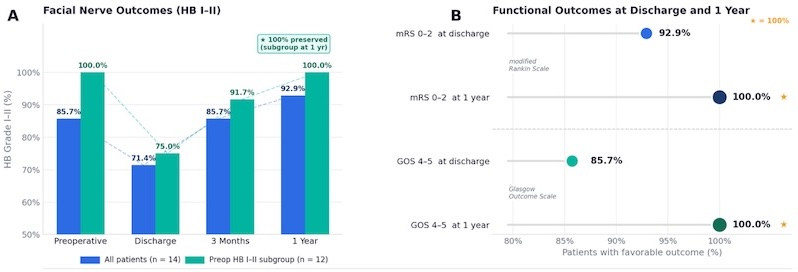
Figure 1. (A) Temporal profile of favorable facial nerve function (House-Brackmann [HB] grade I-II) following translabyrinthine resection of posterior petrous surface meningioma. Rates are shown for all patients (n = 14; blue bars) and the preoperative HB I-II subgroup (n = 12; green bars) at four time points. A transient decline at discharge was followed by progressive recovery; all 12 patients in the preoperative HB I-II subgroup maintained favorable facial nerve function at 1 year (100%). (B) Favorable functional outcomes (MRS 0-2 and GOS 4-5) at discharge and 1 year. Stars (*) denote 100% rates. Dashed line separates the mRS and GOS outcome groups GOS = Glasgow Outcome Scale (favorable = 4-5); HB = House-Brackmann grading scale; HB I-II = favorable (normal to mild paresis); MRS = modified Rankin Scale (favorable = 0-2); preop = preoperative.
Fa, Lin
Gross-Total Resection Versus Function-Preserving Resection With Selective Postoperative Irradiation for Petroclival Meningioma: A Systematic Review and Meta-Analysis
Fa Lin, Md
Department of Neurosurgery, Beijing Tiantan Hospital, Capital Medical University, Beijing, China
Background
For petroclival meningioma (PCM), pursuing gross-total resection (GTR) may improve tumor control but can threaten cranial nerve and brainstem function. Function-preserving non-GTR followed by stereotactic radiosurgery or radiotherapy is increasingly used, yet its comparative effectiveness has not been synthesized separately from reviews of surgical morbidity or radiosurgery outcomes.
Methods
PubMed and Europe PMC were searched from inception to July 28, 2026. Comparative studies of adults with PCM reporting recurrence or progression or functional outcomes according to resection extent or postoperative irradiation were eligible. Risk of bias was assessed using ROBINS-I. Unadjusted recurrence or progression odds ratios (ORs) were pooled using a Paule-Mandel random-effects model with modified Hartung-Knapp confidence intervals. Overlapping cohorts were not double-counted. Certainty of evidence was appraised using GRADE.
Results
Thirteen retrospective cohorts reported 1,575 participants, and five non-overlapping cohorts comprising 670 participants contributed to the meta-analysis. Recurrence or progression occurred in 27 of 288 patients undergoing GTR and 76 of 382 patients undergoing non-GTR. GTR was associated with lower recurrence or progression (OR, 0.26; 95% CI, 0.12-0.56; I2 = 7.9%; 95% prediction interval, 0.09-0.76). The result was similar when partial-resection events were excluded where separable (OR, 0.28; 95% CI, 0.13-0.58), but became imprecise after excluding one study in which events were reconstructed from reported percentages (OR, 0.30; 95% CI, 0.08-1.11). Comparative long-term cohorts suggested that non-GTR followed by postoperative irradiation can approach the tumor control achieved with GTR and outperform non-GTR followed by observation. However, treatment allocation was strongly confounded by anatomy, residual tumor volume, tumor grade, and treatment era. Functional findings were inconsistent and incompletely standardized. Overall certainty of evidence was very low.
Conclusions
GTR is associated with more durable tumor control when it can be performed safely. For tumors with cavernous sinus invasion, brainstem adherence, or neurovascular encasement, planned near-total or subtotal resection with selective postoperative irradiation is a defensible function-preserving strategy. Current evidence does not establish that immediate irradiation is superior to MRI surveillance for every small residual WHO grade 1 PCM. Prospective, anatomy-stratified comparative studies incorporating patient-reported outcomes are required.
Fiorino, Michael
Evolution of Surgical Philosophy in Olfactory Groove Meningioma—A Bibliometric Analysis
Michael Fiorino, Jeroen Coppens
Saint Louis University Hospital
Introduction/Background
The treatment paradigm of olfactory groove meningioma (OGM) has evolved. The advent of endoscopic and minimally invasive surgical approaches has heavily influenced a shift in publication density toward studying these approaches and their potential for functional preservation in OGM; however, the evolution of research priorities within the OGM literature has not been systematically evaluated.
Objectives
To characterize research trends over the last quarter century in the surgical management of OGMs using bibliometric analysis.
Methods/Description
A PubMed search identified English-language publications on OGM management published between 2000 and 2026. Following title and abstract screening, bibliometric analyses were performed using VOSviewer. Keyword co-occurrence, co-authorship, and overlay visualization analyses identified major publication themes, author collaboration networks, and temporal changes in research focus.
Results/Comments
A total of 540 publications met inclusion criteria. The keyword co-occurrence analysis identified four overarching domains: (1) microsurgical anatomy and transcranial approaches, (2) minimally invasive and endoscopic techniques, (3) clinical outcomes and longitudinal follow-up, and (4) oncologic outcomes including recurrence and adjuvant therapy. Overlay visualization demonstrated a temporal shift from publications focusing on operative anatomy and transcranial exposure to those emphasizing endoscopic skull base surgery, anterior cranial base reconstruction, olfactory preservation, quality-of-life outcomes, and individualized patient selection.
Conclusion
This systematic analysis of OGM publications demonstrates an evolution in research priorities over the past two decades. There has been a clear shift toward investigations emphasizing minimally invasive techniques and functional outcomes. These findings serve to illustrate the maturation of OGM research toward individually tailored approaches and patient-centered functional outcomes.
Fletcher, Michael
The History of the Cavernous Sinus: From Anatomical Discovery to Modern Skull Base Surgery
Michael Fletcher, Aysu İyigün Kabakcı, Joao Paulo Almeida
Indiana University School of Medicine
Introduction/Background
The cavernous sinus represents one of the most anatomically and surgically complex regions of the cranium. Progress in the surgical management of cavernous sinus pathologies was hindered for centuries. This stagnation was due to a lack of anatomical understanding of the relationship between the cavernous venous plexus and cranial nerves.
Objectives
To review the history of the cavernous sinus, from conception to the modern era, and examine how historical anatomical discoveries have driven surgical innovation.
Methods/Description
A literature review was conducted utilizing PubMed, Google Scholar, and primary historical texts. Included studies consisted of anatomical studies, surgical reports, and historical papers, which were reviewed and synthesized chronologically.
Results/Comments
The earliest observations by Claudius Galen and Raymond de Vieussens laid the anatomical framework of the parasellar region. Following these discoveries, surgical stagnation occurred for nearly fifteen centuries. In the eighteenth century, Jacques Bénigne Winslow popularized the term cavernous sinus because of its resemblance to the corpora cavernosa. It was not until the twentieth century that George Parkinson reported the first successful direct surgical treatment of a carotid-cavernous fistula, demonstrating surgical feasibility. Vinko Dolenc subsequently developed reproducible extradural skull base approaches. Finally, Albert Rhoton meticulously characterized the microsurgical anatomy of the cavernous sinus, enabling the development of modern skull base surgery.
Conclusion
The evolution of cavernous sinus surgery illustrates the relationship between anatomical studies and surgical innovation. The pioneering anatomical work of Rhoton, coupled with surgical innovations of Parkinson and Dolenc, transformed the cavernous sinus from an inoperable region to a cornerstone of modern skull base surgery.
Conflict of Interest Disclosure
The authors declare no conflicts of interest related to this work.
Funding
A. İyigün Kabakcı received scholarship support from the Turkish Neurosurgical Society during the conduct of this work.
Gabbita, Abhirama C.
At the Crossroads of Petrous and Tentorium: A Novel Classification and Surgical Series of Petro-Tentorial Meningiomas via the Retromastoid Approach
Abhirama Chandra Gabbita, Subodh Raju, Suresh Kanasani
AIG
Introduction: Petro-tentorial meningiomas, arising at the junction of the petrous ridge and tentorial leaflet, represent an uncommon and anatomically distinct subset of posterior fossa meningiomas. Unlike petroclival or purely tentorial lesions, these tumors possess a unique dural attachment spanning the petrous and tentorial surfaces, often involving critical neurovascular structures including cranial nerves V–XI, the superior petrosal venous complex, and adjacent dural sinuses. No dedicated classification currently exists to guide surgical planning for these lesions through the retromastoid supracerebellar-infratentorial (RSCIT) corridor.
Methods: We retrospectively reviewed six consecutive patients with petro-tentorial meningiomas treated via a retromastoid approach. Based on preoperative imaging, dural attachment, compartmental extension, and neurovascular relationships, we propose a hybrid anatomo-radiological classification adapted from established petroclival and tentorial meningioma systems: Type I (posterior petro-tentorial), Type II (medial/petroclival), and Type III (bi-compartmental/trans-tentorial).
Results: The cohort was predominantly female (83.3%) with a mean age of 48.3 ± 7.2 years. Headache (83%) and ataxia (67%) were the most common presenting symptoms. Gross total resection (Simpson Grade I–II) was achieved in 83.3% of cases. One patient (16.7%) developed transient postoperative cranial neuropathy, which improved during follow-up. No cerebrospinal fluid leaks, major vascular injuries, or perioperative mortality were observed. Mean Karnofsky Performance Status improved significantly from 80 ± 5 preoperatively to 90 ± 6 postoperatively (p = 0.03). No tumor recurrence was identified during a mean follow-up period of 24.5 months.
Conclusion: The retromastoid supracerebellar-infratentorial approach enables maximal safe resection of selected petro-tentorial meningiomas while preserving neurological function. The proposed Type I–III classification provides a practical, corridor-oriented framework for preoperative planning, surgical strategy, and standardized reporting, and warrants validation in larger multicenter studies.
Galindo Rico, Christian Emmanuel
Radiotherapy-Induced Histopathological Changes Associated with Reduced Intraoperative Bleeding in an Intraventricular Meningioma: A Case Report
Diana María Silva-Romero, Karina Amezcua-Barragán, Sergio Moreno-Jiménez, Karely Valencia-Guzmán, Karen Alejandra Miranda-Fernández, Fabiola Flores-Vázquez, Isaac Vargas-Olmos, Martha Lilia Tena-Suck, Julieta Vázquez-García, Christian Emmanuel Galindo Rico
National Institute of Neurology and Neurosurgery ‘Manuel Velasco Suárez’, Mexico City, Mexico
Introduction/Background
Hypervascular meningiomas remain surgically challenging because excessive intraoperative bleeding may compromise the safety and extent of resection. Although preoperative embolization has been extensively investigated, the histopathological effects of preoperative radiotherapy on tumor vascularity and surgical resectability remain poorly characterized.
Objectives
To describe the histopathological changes induced by preoperative radiotherapy in a giant intraventricular meningioma and explore their association with reduced intraoperative bleeding and improved surgical handling.
Methods/Description
A 30-year-old woman presented with intracranial hypertension and progressive left hemiparesis caused by a giant right intraventricular meningioma. Initial subtotal resection was interrupted because of massive intraoperative hemorrhage (1,500 mL). Histopathological examination demonstrated a WHO CNS grade 1 meningothelial meningioma. Following stereotactic fractionated radiotherapy (54 Gy in 30 fractions), tumor progression necessitated repeat surgery nine months later. Histopathological specimens obtained before and after radiotherapy were comparatively analyzed.
Results/Comments
Repeat resection was performed with substantially reduced blood loss (400 mL). Intraoperatively, the tumor appeared paler, firmer, less vascular, and exhibited a more favorable dissection plane. Comparative histopathological analysis demonstrated increased stromal fibrosis, collagen deposition, vascular wall thickening, edema, necrosis, reduced cellularity, and markedly increased type IV collagen expression following radiotherapy. Ki-67 increased from 1% to 8%, while the tumor retained the histopathological features of a WHO CNS grade 1 meningothelial meningioma.
Conclusion
This case provides histopathological evidence that preoperative radiotherapy may induce vascular and stromal remodeling associated with reduced intraoperative bleeding and improved surgical handling in giant hypervascular meningiomas. These findings support further investigation of preoperative radiotherapy as a potential adjunctive strategy in selected complex meningioma cases.
Gendle, Chandrashekhar
EXOSCOPE-ENDOSCOPE-ASSISTED MINIMAL INVASIVE REMOVAL OF LATERAL VENTRICULAR SUB- EPENDYMAL GIANT CELL ASTROCYTOMA
Dr chandrashekhar Gendle, Dr.Sivashanmugam Dhandapani, Dr. Prachi Gang
PGIMER
Background
SEGAs are slow growing, WHO grade 1 tumours often arising near the foramen of monro within the lateral ventricles. In this paper, we present a case of a 55year old lady who presented to us with isolated, progressively worsening, episodic headaches since 1 year with no other positive history or relevant examination findings. She was managed via a minimally invasive transcortical approach utilizing combined exoscopic and endoscopic visualisation to achieve safe and effective tumour removal with minimal cortical disruption.
Methods/technique
She was positioned in right supine lateral position and a linear incision was marked with Neuronavigation guidance in the mid 1/3rd parasaggital area. A Rt parasaggital craniotomy was made and dura was reflected over the SSS. Dissection was advanced via an Interhemispheric approach and b/l pericallosal arteries were identified and safeguarded. Targeted Callosotomy was performed after confirmation on navigation. The tumour was greyish, soft, partly suckable, and cusable. NTR done. Check endoscopy towards the end of resection helped identify hidden slivers of tumour tissue, which were then carefully excised.
Results/outcomes
She was extubated, clinically stable with no new deficits. Her post op scan was unremarkable. Her final biopsy report was s/o SEGA.
Conclusion
The use of advanced visualisation tools such as exoscopes and endoscopes had enhanced surgical precision by improving depth perception, illumination , and ergonomics in deep seated lesions.
Gendle, Chandrashekhar
Unilateral Extended Frontal Craniotomy for Midline Anterior Skull Base Meningiomas: Preserving Olfaction and Minimising Surgical Morbidity
Dr Chandrashekhar Gendle, Dr Pravin Salunke, Dr Madhivanan Karthigeyan, Dr Keshav Mishra
PGIMER, CHANDIGARH,INDIA
Abstract
Background
Resecting anterior cranial fossa(ACF) meningiomas remains surgically demanding due to their deep midline location and intimate relationships with the olfactory tracts and the superior sagittal sinus (SSS). Conventional extended bifrontal craniotomies, while providing generous exposure, frequently necessitate excessive brain retraction, risk inadvertent injury to neurovascular bundles, require SSS ligation, and are associated with a disturbingly high incidence of new-onset anosmia, a complication that profoundly diminishes postoperative quality of life. To mitigate these inherent drawbacks, we developed and implemented a unilateral extended frontal approach.
Objective
This study evaluates the safety and efficacy of this novel technique, specifically assessing its capacity to minimise cerebral manipulation, preserve SSS integrity, and optimise functional olfaction without compromising the radicality of tumour resection.
Methods
Thirteen patients with ACF meningioma were operated on using the novel technique of unilateral extended frontal skull base approach. The clinical presentation, radiological studies, intraoperative findings, and outcome at follow-up were recorded..
Results
Gross total tumour resection was achieved in 12 of 13 patients. At least one olfactory tract was anatomically preserved in all patients, and SSS was preserved in all patients. Functional olfaction preservation was achieved in 8 patients. No patient developed new-onset anosmia.
Conclusion
The extended unilateral frontal approach is a viable and reliable alternative for the extended bifrontal technique for the resection of large midline ACF meningiomas with avoidance of SSS ligation, decreased brain handling with better olfaction preservation, while achieving comparable tumour resection and acceptable cosmetic outcomes
Gopeesingh, Neil
Paediatric Dural-based Spindle Cell Neoplasm: A diagnostic dilemma
Neil Gopeesingh, Priyadashni Ramsamooj, Uppalapati Kali, Ramcharan Robert
Port of Spain General Hospital – Trinidad and Tobago
Introduction/Background
Meningiomas are the most common primary intracranial tumours but are rare in the paediatric population. Diagnostic challenges arises when histopathological and immunohistochemical findings overlap with other spindle cell neoplasms. We present a rare occipital dural-based spindle-cell neoplasm in an adolescent with histological features suggestive of both fibrous meningioma and a schwannoma.
Objectives
To describe the clinical, radiological, operative and pathological features of a paediatric of an occipital spindle-cell neoplasm with overlapping features of fibrous meningioma and schwannoma.
Methods/Description
A 16-year-old female presented with a right occipital mass. MRI demonstrated a 4.7 cm × 3.4 cm × 3.21 cm well-defined extra-axial lesion with heterogeneous signal characteristics and necrotic core with surrounding vasogenic edema. The lesion was surgically resected and submitted for histopathological and immunohistochemical analysis.
Results/Comments
Intraoperatively, the tumour was firm, extra-axial, supratentorial and dural-based, abutting the tentorium cerebelli and falx cerebri. Histology demonstrated a spindle-cell neoplasm composed of fascicles and bundles of elongated cells with focal nuclear pleomorphism. Immunohistochemistry revealed uniform S100 positivity, variable SOX10 expression, focal EMA positivity, variable GFAP expression, and variable SSTR2A staining. Despite the ambiguous immunophenotype, correlation of radiological and operative findings, together with the rarity of supratentorial dural-based schwannomas supported a diagnosis of fibrous meningioma.
Conclusion
This case highlights the importance of multidisciplinary clinico-radiological and pathological correlation in accurately classifying rare paediatric dural based spindle cell neoplasms. Recognition of such atypical presentations is crucial for appropriate diagnosis and subsequent management.
Gött, Hanna
Characterizing Radiation-Induced Meningiomas: Epidemiological, Histopathological, and Clinical Findings from 52 Cases
Hanna Goett, Hannes Becker, Jonas Tellermann, Christina Fodi, Felix Behling, Marcos Tatagiba
Department of Neurosurgery, University Hospital Tuebingen
Introduction/Background
Radiation-induced Meningioma (RIM) are a rare but clinically relevant long-term sequelae of ionizing irradiation applied to the central nervous system. They can be distinguished from spontaneous meningioma on a clinical and histomolecular basis. Due to their low frequency little is known about their clinicopathological characteristics.
Objectives
We present a series of 52 cases of RIMs to highlight the specific epidemiological and histopathological characteristics and clinical course.
Methods/Description
In a total cohort of 3159 cases of surgical treatment for meningioma between 2004 and 2022 we discovered 52 RIM from 26 patients which had prior radiation. Extend of resection according to Simpson grade was taken from surgical reports. Further clinical factors were obtained from clinical records.
Results/Comments
Mean age at diagnosis was 47.1 (SD 11.9) years with a mean latency of 31.5 years (SD 14.0). We observed male predominance with n=33 (63.5%) compared to n=21 (36.5%) female patients. Only 20 RIM were located in the skull base while 32 cases could be attributed to the convexity (n=23) and falx (n=9) region. 59.6% of RIM were classified as WHO Grade 2 and 3, while only 41.4% represented WHO Grade 1 and histopathological brain invasion was pronounced in 20% of RIM. Meningiomatosis was present in 25.5% of cases. Median PFS was 63.4 months.
Conclusion
RIMs demonstrated frequent aggressive features, underscoring their distinct biological behavior and the need for long-term clinical surveillance.
Gött, Hanna
Pediatric Meningiomas Differ from Adult Disease: Insights from a 42-Patient Single-Center Cohort
Hanna Goett, Hannes Becker, Jonas Tellermann, Christina Fodi, Felix Behling, Martin Schuhmann
Department of Neurosurgery, University Hospital Tuebingen, Eberhard Karls University Tuebingen
Introduction/Background
Pediatric meningiomas are rare tumors, accounting for only a small proportion of CNS neoplasms in children. Their low incidence has limited the availability of comprehensive data regarding presentation, management, and outcomes.
Objectives
To characterize the clinical and histopathological features of pediatric meningiomas and to evaluate treatment outcomes in a large single-center cohort.
Methods/Description
We retrospectively analyzed 42 patients aged 3–18 years who underwent surgery for meningioma at a tertiary high-volume neurosurgical center between 2004 and 2022. Complete clinical and imaging follow-up data were available for all patients. Epidemiological, clinical, radiological, and histopathological parameters were extracted from medical records. Tumor recurrence was defined by MRI imaging.
Results
The mean age at diagnosis was 13.3 years (95% CI: 12.2–14.5). Multiple meningiomas were present in 40.5% of patients, and 64.3% had confirmed Neurofibromatosis Type 2 (NF2). Previous radiotherapy for another malignancy was documented in two cases. Tumors were located in the spine in 9.5% and intraventricularly in 7.1% of patients. WHO grade 2 and 3 tumors accounted for 23.7% of cases. The mean MIB-1 proliferation index was 2.9%. Tumor recurrence occurred in 28.6% of patients, with a mean progression-free survival (PFS) of 44.7 months (95% CI: 27.9–61.5). Sporadic cases showed a shorter PFS than NF2-associated tumors, although this difference was not statistically significant.
Conclusion
Pediatric meningiomas demonstrate distinct characteristics compared with adult meningiomas, including a higher prevalence of multiple tumors, NF2 association, and recurrence. These findings highlight the need for multidisciplinary management and long-term surveillance in affected patients.
Gray, Landon
Surgical strategies for olfactory preservation in olfactory groove meningioma: a systematic review and technical recommendations
Landon Gray, Alice Giotta Lucifero, M.d., PhD, Estefana Bcharah, B.s., Matthew Dufault, Amirhossein Akhavan-Sigari, Bernard Bendok, M.s.c.i.
Mayo Clinic
Introduction/Background
Olfactory dysfunction is a common post-operative complication in patients with olfactory groove meningiomas (OGMs). Although gross-total resection remains the primary goal, the factors influencing postoperative olfactory preservation remain incompletely characterized.
Objectives
To synthesize evidence regarding olfactory preservation following OGM resection, identify surgical factors associated with favorable outcomes, and develop technical recommendations to guide surgical decision-making.
Methods/Description
A systematic review of literature was performed. Studies reporting surgical treatment of OGMs with olfactory outcomes were reviewed. Extracted variables included preoperative olfactory status, tumor characteristics, surgical approach, olfactory outcomes, and preservation strategies.
Results/Comments
Twelve studies comprising 192 patients were included. Olfactory preservation was achievable with transcranial and selected endoscopic approaches, particularly in patients with intact preoperative olfaction, small unilateral tumors, limited cribriform involvement, and no significant midline extension. Preservation of a single olfactory pathway was often sufficient to maintain postoperative function. Key technical strategies included preserving at least one olfactory bulb and tract, maintaining arachnoid planes, minimizing frontal lobe retraction, limiting coagulation near the olfactory apparatus, and using unilateral corridors when feasible. Several studies reported olfactory preservation rates exceeding 60% with olfaction-sparing transcranial approaches, whereas extensive bilateral tumors, complete cribriform involvement, and preoperative anosmia were consistently associated with poor outcomes.
Conclusion
Olfaction can often be preserved in carefully selected patients through olfaction-sparing surgical techniques, with favorable outcomes primarily determined by tumor anatomy, preoperative olfactory function, and preservation of at least one olfactory pathway.
Guerrero, Miguel
When Imaging Reveals More Than Expected: An Incidental Falcine Meningioma
Miguel Guerrero, Ricardo Y. Topete, Mario Messina, Yamil O. Ochoa, Natalia Macias, Fernanda Lopez, Citlalli S. Guerrero
Hospital Civil Juan I Menchaca
Background
Meningiomas are the most common benign primary tumors of the central nervous system, accounting for approximately 36.4% of intracranial neoplasms. Falcine meningiomas arise from the falx cerebri and may extend bilaterally, often remaining clinically silent until they reach a considerable size.
Objective
To describe the clinical presentation, imaging findings, surgical management, and outcome of a patient with a falcine meningioma.
Methods
Retrospective review of clinical records, neurological examination, neuroimaging studies, operative reports, histopathological findings, and postoperative follow-up. Case Presentation: An elderly male presented after a ground-level fall caused by sudden bilateral lower-extremity weakness. Neurological evaluation revealed no loss of consciousness or seizures. CT and MRI demonstrated a large extra-axial solid-cystic lesion with marked perilesional edema, consistent with a falcine meningioma. The patient underwent a right precoronal craniotomy using an interhemispheric approach. Gross total resection was achieved through microsurgical devascularization, tumor debulking, and resection of the involved falx while preserving both anterior cerebral arteries.
Results
Intraoperatively, the lesion demonstrated bilateral falcine extension and involvement of both anterior cerebral arteries. Postoperatively, the patient developed transient stupor and disorientation (GCS 13) but showed progressive neurological improvement. Follow-up CT revealed minimal pneumocephalus without significant mass effect. At discharge, he was alert, coherent, tolerating oral intake, and had only mild residual temporospatial disorientation.
Conclusion
Surgical resection remains the cornerstone of treatment for falcine meningiomas. Careful microsurgical technique can achieve favorable outcomes even in complex lesions involving critical vascular structures.
Gump, William
The Atypical is Typical: Pediatric Meningiomas and Mimics
William Gump
Norton Children’s Hospital
Meningioma is an atypical pathology in pediatrics. A single-surgeon experience over 15 years is reviewed. A total of 4 cases of meningioma were identified, all in patients 16 or 17 years old. Of these patients, 2 had radiographic features typical of meningioma, including a patient with a history of radiation therapy for Hodgkin lymphoma. In the other 2 cases meningioma was not considered on the differential diagnosis preoperatively. 3 cases were grade 1 pathology, and 1 case was grade 2. Each patient had a histologic subtype unique to the cohort. In addition, 4 other patients ranging from 4 to 9 years of age were identified in which meningioma was considered the most likely diagnosis preoperatively, based on imaging features, but were found to have other diagnoses from tissue analysis.
Diagnoses mimicking meningioma on imaging included Rosai-Dorfman disease, myeloid sarcoma, embryonal rhabdomyosarcoma, and glioma with astrocytic features and BANP-ALK rearrangement. The Rosai-Dorman lesion was in the ventral cervical spinal canal and was initially diagnosed as meningioma from histopathology prior to methylation profiling; all other patients had intracranial lesions. One meningioma was purely intraventricular. The 4 meningioma patients represented only 1.5% out of a total 260 unique patients with brain tumors who were treated over the study interval. There were no meningioma cases among the 32 patients in the study period with intradural spinal neoplasms. Although the small number of patients in this cohort precludes drawing statistical conclusions, this study illustrates the scarcity and heterogeneity of meningioma presentation in children.
Ihata, Tomohiro
Surgical Strategy for Recurrent Meningioma with Orbital Invasion: A Case Report and Literature Review
Tomohiro Ihata, Ryokichi Yagi, Hideki Kashiwagi, Yoshinari Oue, Masao Fukumura, Gen Futamura, Motomasa Furuse, Masahiko Wanibuchi
Osaka Medical and Pharmaceutical University
Background
Recurrent meningiomas with orbital invasion often involve the optic nerve, orbital apex, and cavernous sinus, making complete resection challenging. Surgical management aims to achieve maximal safe resection while preserving visual and neurological function.
Objective
To describe our surgical strategy for recurrent meningioma with orbital invasion and discuss the rationale for surgical approach selection.
Case Description
A man in his 30s presented with progressive right-sided proptosis six years after resection of a right middle cranial fossa meningioma. Magnetic resonance imaging demonstrated recurrent tumor extending through the superior orbital fissure into the orbit and contacting the lateral rectus muscle. Visual acuity, visual fields, and ocular motility were preserved. Because extensive postoperative adhesions and orbital extension were anticipated, a transcranial orbital approach was selected. Microsurgical dissection enabled safe tumor removal while preserving the optic nerve and surrounding neurovascular structures. Subtotal resection was intentionally performed to preserve visual function, followed by planned adjuvant radiotherapy.
Discussion
Selection of the surgical approach should be individualized according to tumor extension, intraconal or extraconal location, relationship to the optic nerve, and history of previous surgery. For recurrent tumors requiring wide field of view, precise microsurgical manipulation, and careful adhesion dissection, the transcranial approach remains advantageous.
Conclusion
A transcranial orbital approach is an effective option for recurrent meningiomas with extensive orbital involvement, enabling maximal safe resection while preserving visual function. Postoperative radiotherapy should be considered when indicated to improve long-term local control.
Januschek, Elke
When Resection Is Not Enough: The Truly Curious Case of Relentless Meningioma Growth — Is There Anything Left to Do?
Elke Januschek, Serine Ajlouni
Sana Klinikum
Background
Simpson grade I resection with excision of the involved dura is regarded as the most effective strategy for durable meningioma control, with recurrence typically attributed to residual local disease. Persistent tumor formation following anatomically complete resections is therefore unexpected and suggests alternative mechanisms of disease development.
Objective
To investigate repeated meningioma formation following multiple confirmed Simpson grade I resections and evaluate whether this pattern represents true recurrence or reflects a non- biological process.
Methods
A 39 -year-old patient presented with the first right frontotemporal meningioma in 2012 and underwent Simpson grade I resection. Subsequent resections of new meningiomas in 2016, 2018, and 2025 resulted in cumulative excision of previously involved dura. Multimodal adjuvant therapies including radiotherapy, immunotherapy, and radionuclide therapy were administered between 2019 and 2024. Clinical course and serial MRI studies were retrospectively analyzed within a multidisciplinary framework.
Results
Serial imaging demonstrated repeated emergence of spatially distinct, non-contiguous meningiomas rather than regrowth at prior operative sites. Notably, all tumors developed exclusively within the right cerebral hemisphere despite anatomically complete resections with cumulative dural excision.
Conclusion
Continued tumor formation despite repeated Simpson grade I resections challenges the assumption that meningioma recurrence is primarily driven by residual local disease. The consistent hemispheric laterality supports a regionally determined biological susceptibility and suggests limitations of purely anatomy-based treatment paradigms.
Kantarci, Kemal
Microsurgical Resection of a C2 Schwannoma by a Unilateral Posterior Cervical Approach Without Bone Resection
Kemal Kantarci, Semih Fidan, Abuzer Güngör, Uğur Türe
MedicalPark Bahvelievler Hospital – Altınbaş University
Introduction/Background
Spinal schwannomas at C1-C2 account for 5% of spinal nerve root tumors. Unlike other intervertebral joints, the C1-C2 region lacks a natural bony neural foramen, leaving the C2 dorsal root ganglion without bony coverage due to upper cervical anatomical differences. Therefore, tumors without major extraspinal or intradural extensions are clearly visible between the C2 lamina and C1 posterior arch.
Objectives
To present an alternative, tissue-sparing surgical technique for the complete resection of primary nerve sheath tumors in the upper cervical spine without laminectomy, capitalizing on this unique regional anatomy.
Methods/Description
We demonstrate the microsurgical resection of a C2 schwannoma in a 20-year-old female with neurofibromatosis type I. A standard posterior midline approach was used with unilateral dissection on the lesion’s side. Because of the distinct C1-C2 anatomy, the resection was performed entirely through natural surgical corridors, without any bone resection.
Results/Comments
This approach provided sufficient anatomical exposure for the successful and complete removal of the tumor. Avoiding bone removal allowed the procedure to be performed in less time while strictly respecting the structural stability of the upper cervical vertebrae.
Conclusion
The unilateral posterior cervical approach without bone resection leverages the unique C1-C2 anatomy. It is a safe, tissue-sparing, and time-efficient alternative for appropriate cases of C2 schwannomas that do not extend intradurally to adjacent segments of the spinal cord.
Kelani, Aminath B.
Cranial electrical injury and meningiomatosis: coincidence or consequence?
Aminath B. Kelani, Karima Mokhtari, Matthieu Peyre, Ali Boubacar, Saïdou Madougou, Philippe Cornu
Faculty of Health Sciences, Abdou Moumouni University, Niamey, Niger
Li, Boyi
Outcomes after surgical resection of tuberculum and planum meningiomas – A single-center, single-surgeon case series
Boyi Li, Zoe Robinow, Carlos David
University of North Carolina
Introduction
Tuberculum and planum meningiomas often present with progressive visual impairment. The optimal surgical approach for resection remains a topic of debate.
Objectives
Here we present a single-center, single-surgeon consecutive case series of outcomes after transcranial approach for surgical resection of tuberculum and planum meningiomas.
Methods
We performed a retrospective review of a single surgeon’s consecutive cases from January 2022 to October 2025, including only tuberculum and planum meningiomas.
Results
25 patients (23 female, 2 male) were included. Average age was 53 years. Average BMI was 34.7. 16 (64%) patients presented with vision loss while 4 (16%) presented with other visual symptoms. Average tumor size was 2.2 cm. 22 (88%) of the tumors directly compressed the optic apparatus, 13 (52%) had sellar involvement, 13 (52%) encased major blood vessels, 8 (32%) invaded the optic canal, and 3 (12%) invaded the cavernous sinus. 18 (72%) patients underwent orbitocranial craniotomies, 4 (16%) underwent pterional craniotomies, and 3 (12%) required orbitozygomatic craniotomies. Average length of stay was 4.2 days, with 24 (96%) patients discharging home. There was no postoperative mortality, hemorrhage, stroke, wound dehiscence, cerebrospinal fluid leak, or infection over average length of follow-up of 281 days. 21 (84%) patients had gross total resection, with no evidence of recurrence on follow-up MRIs. Overall, 23 (92%) patients had favorable visual outcomes based on preoperative and postoperative ophthalmologic testing.
Conclusions
We demonstrate the reliability and efficacy of the transcranial approach in experienced hands for surgical resection of tuberculum and planum meningiomas, with respect to achieving optimal tumor resection and preservation of vision.
Lucifero, Alice Giotta
Comparative Anatomical Study of Transbasal Approaches: Suprasinus, Transsinus, and Orbital Rim Removal Variants Using High-Definition 3D Photogrammetry
Alice Giotta Lucifero, Evelyn Turcotte, Matthew Dufault, Amirhossein Akhavan-Sigari, Vita Olson, Estefana Bcharah, Landon Gray, Alexandre Bossi Todeschini, Charbel Moussalem, Bernard Bendok
Mayo Clinic
Introduction
The transbasal approach remains a cornerstone of skull base surgery, providing a direct midline corridor to the anterior cranial fossa. Over time, several modifications have been introduced to expand surgical exposure and minimize frontal lobe retraction. Among these, the suprasinus, transsinus, and orbital rim removal variants are predominant in clinical practice, each with anatomical advantages and limitations.
Objectives
To compare the surgical exposure, operative trajectories, working angles, and anatomical constraints of three transbasal approaches.
Methods
Five formalin-fixed, latex-injected adult cadaveric heads underwent sequential transbasal dissections. A suprasinus, transsinus, and orbital rim removal were performed in a stepwise fashion. High-definition 3D photogrammetry models were generated throughout the dissections for comparative analysis of surgical freedom, working angles, exposure, and anatomical constraints.
Results
The suprasinus approach provided direct access to the anterior cranial fossa while preserving the frontal sinus. Dural opening could be performed unilaterally or bilaterally depending on the extent of the lesion, although wider exposure frequently required division of the falx. Opening the frontal sinus through the transsinus approach shortened the surgical trajectory and improved visualization of the cribriform plate, planum sphenoidale, and central skull base. The addition of orbital rim removal further lowered the operative corridor, significantly increasing inferior-to-superior viewing angles and reducing the need for frontal lobe retraction.
Conclusion
Progressive modifications of the transbasal approach improve exposure, and surgical freedom. High-definition 3D photogrammetry enables detailed visualization of these anatomical differences and supports a practical framework for tailoring the transbasal approach according to lesion location and surgical objectives.
Lucifero, Alice Giotta
Fissural Corridors of the Posterior Fossa: A Skull Base Anatomical Study Integrating 3D Photogrammetry Toward an Approach-Selection Algorithm.
Alice Giotta Lucifero, Alexandre Bossi Todeschini, Estefana Bcharah, Matthew Dufault, Landon Gray, Maria José Pachón-Londoño, Amirhossein Akhavan-Sigari, Evelyn Turcotte, Charbel Moussalem, Bernard Bendok
Mayo Clinic
Introduction
Selecting the optimal approach for posterior fossa meningiomas remains challenging. Cerebellar-brainstem fissures and cisternal spaces serve as key surgical corridors that expand operative windows, minimizing morbidity and maximizing exposure.
Objectives
This study aimed to define the anatomical advantages and limitations of major posterior fossa approaches and fissural corridors to develop a practical approach-selection framework.
Methods
Five formalin-fixed adult cadaveric heads underwent systematic microsurgical dissection. High-definition 3D photogrammetric reconstruction was performed to generate spatial models for analyzing surgical trajectories, exposure, and anatomical constraints. Representative clinical cases were used to validate the proposed algorithm.
Results/Comments
On the tentorial surface, the supracerebellar infratentorial, posterior interhemispheric, and suboccipital transtentorial approaches utilized the cerebellomesencephalic and primary fissures to access the upper fourth ventricle, upper pons, and middle cerebellar peduncle. On the suboccipital surface, the midline suboccipital approach with tonsillouvular fissure splitting widened access to the fourth ventricle, while the far-lateral approach expanded exposure through the cerebellomedullary fissure to the caudal fourth ventricle and cerebellopontine angle. On the petrosal surface, the retrosigmoid approach and medial/lateral petrosal fissure splitting enabled access to the cerebellopontine angle and lateral pons. A stepwise algorithm integrating surface anatomy, fissure use, approach selection, and target definition was developed.
Conclusions
Posterior fossa fissures constitute natural skull base corridors that can be systematically used for individualized surgical planning. The integration of microsurgical anatomy, 3D photogrammetry, and illustrative cases enables the development of a practical approach-selection algorithm, enhancing precision and safety in posterior fossa surgery.
Lucifero, Alice Giotta
Microsurgical Optic Canal Unroofing in Skull Base Meningioma Surgery: Anatomical Principles and Technical Nuances
Alice Giotta Lucifero, Charbel Moussalem, Matthew Dufault, Amirhossein Akhavan-Sigari, Maria Josè Pachón-Londoño, Evelyn Turcotte, Estefana Bcharah, Landon Gray, Alexandre Bossi Todeschini, Bernard Bendok
Mayo Clinic
Introduction
Optic canal invasion is frequently found in clinoidal, parasellar, and spheno-orbital meningiomas and represents a major cause of progressive visual deterioration. Early decompression of the optic nerve is essential for preserving visual function while maximizing tumor resection.
Objectives
This study illustrates the anatomical principles, technical nuances, and surgical relevance of optic canal unroofing in the treatment of skull base meningiomas.
Methods
Patients underwent resection of skull base meningiomas with evidence of optic canal involvement were retrospectively reviewed. Surgical videos and operative records were analyzed to identify key anatomical landmarks, technical steps, and operative strategies.
Results
Pterional and cranio-orbitozygomatic approaches provided wide exposure of the optic canal, internal carotid artery, and oculomotor nerve. The falciform ligament was identified and incised, exposing the optic roof, which was subsequently drilled with a high-speed diamond burr under continuous irrigation. Drilling can be tailored and further extended to the medial or lateral compartemtn of the anterior clinoid process. Unroofing of the optic canal allowed optic nerve decompression and facilitated tumor dissection and gross-total resection when anatomically feasible. Visual function remained stable or improved in the majority of patients, with low rates of procedure-related morbidity.
Conclusion
Optic canal unroofing represents a critical step in the microsurgical management of skull base meningiomas. This technique expands operative exposure, provides early optic nerve decompression, and facilitates maximal tumor resection while preserving or restoring visual function.
Lucifero, Alice Giotta
Stepwise Microsurgical Unroofing of the Optic Canal: An Anatomical Study with High-Definition 3D Photogrammetry
Alice Giotta Lucifero, Mattew Dufault, Amirhossein Akhavan-Sigari, Vita Olson, Evelyn Turcotte, Estefana Bcharah, Landon Gray, Alexandre Bossi Todeschini, Charbel Moussalem, Bernard Bendok
Mayo Clinic
Introduction
The optic canal is a critical anatomical landmark for lesions involving the parasellar and anterior skull base regions. Optic canal unroofing is a fundamental surgical maneuver that requires a thorough understanding of the anatomy and microsurgical technique.
Objectives
To review the microsurgical anatomy of the optic canal and develop a reproducible step-by-step technique for optic canal unroofing using cadaveric dissection supplemented by high-definition 3D photogrammetry.
Methods
Three formalin-fixed, latex-injected cadaveric heads underwent step-by-step microsurgical dissections of optic canal unroofing. Stages of the procedure were documented using high-resolution three-dimensional models. Surgical landmarks, drilling boundaries, neurovascular relationships, and operative corridors were systematically analyzed.
Results
Following exposure of the optic canal, the falciform ligament overlying the canal was identified and mobilized. The ligament was then incised, and the dural fold reflected inferiorly to expose the underlying bony canal. The medial wall of the canal was progressively drilled using a high-speed diamond burr under continuous irrigation. When additional lateral exposure was required, drilling was extended toward the medial or lateral aspect of the anterior clinoid process and tailored according to the extent and location of the lesion. Three-dimensional reconstructions accurately demonstrated the spatial relationships among the neurovascular structures, enabling precise identification of critical landmarks and safe drilling boundaries.
Conclusions
Microsurgical optic canal unroofing can be standardized through a sequence of anatomical steps that facilitate safe decompression of the optic nerve. The integration of cadaveric dissection and 3D photogrammetry provides a valuable educational and surgical planning tool, facilitating the translation of anatomical knowledge into operative practice.
Lucifero, Alice Giotta
The Emerging Role of Liquid Biopsies in Meningioma Diagnosis, Prognostication, and Precision Management: A Systematic Review
Zeinab Hammoud, Amirhossein Akhavan-Sigari, Alice Giotta Lucifero, Matthew Dufault, Landon Gray, Mytreyi Trivedi, Estefana Bcharah, Bernard R. Bendok
Lebanese University
Introduction
Molecular profiling has expanded our understanding of meningioma biology and created opportunities for improved diagnosis, prognostication, and personalized treatment. Liquid biopsy approaches, including circulating nucleic acids, extracellular vesicles, and other molecular biomarkers, offer a minimally invasive method for assessing tumor biology. We performed a systematic review to evaluate the current evidence supporting liquid biopsy applications in meningioma management.
Methods
A systematic review was conducted following PRISMA guidelines. PubMed/MEDLINE, Embase, Web of Science, and Cochrane CENTRAL were searched. Original studies evaluating liquid biopsy biomarkers in patients with intracranial meningiomas were included. Outcomes of interest included diagnostic performance, tumor classification, prognostication, recurrence prediction, treatment monitoring, and potential utility for precision therapies. Due to anticipated heterogeneity in biomarker platforms and outcome measures, findings were synthesized qualitatively.
Results
Current evidence suggests that liquid biopsy biomarkers may assist in meningioma detection, molecular characterization, grading, and prediction of clinical behavior. However, translation into routine clinical decision-making remains limited by small sample sizes, heterogeneous methodologies, and lack of prospective validation.
Conclusion
Liquid biopsy represents a promising avenue for minimally invasive molecular assessment of meningiomas. Further standardized and prospective studies are needed to establish clinical utility and facilitate integration into personalized meningioma care.
Malazonia, Zviad
Microsurgical Management of Medial Sphenoid Wing Meningiomas: A Retrospective Single-Center Study
Zviad Malazonia, Vladimer Tsikarishvili, Mirza Khinikadze, Archil Agladze, Tornike Kldiashvili, Rudiko Jigania, Nano Chochia
Caucasus International University
Background
Medial sphenoid wing meningiomas (MSWMs) are among the most challenging skull base tumors because of their intimate relationship with the cavernous sinus, optic apparatus, and major intracranial vessels.
Objectives
To evaluate clinical presentation, surgical strategy, and postoperative outcomes in microsurgically managed MSWMs.
Methods
We retrospectively reviewed 52 patients with MSWMs treated between January 2018 and June 2026. Microsurgical resection was performed using pterional or orbitozygomatic approaches, frequently combined with extradural anterior clinoidectomy. The extent of resection was assessed using the Simpson grading system.
Results
The cohort included 52 patients with a median age of 48 years. Gross-total resection in Simpson Grade I was achieved in 30 patients (57.7%), and Simpson Grades II–III in 12 patients (23.1%), while subtotal resection (Grade IV) was performed in 10 patients (19.2%), leaving residual tumor due to cavernous sinus invasion, adherence to the neurovascular structures, and optic canal involvement. CSF leaks occurred in 8 patients (15.3%), cranial nerve (CN) III palsy developed in 8 patients (15.3%), which was transient in 6 patients and permanent in 2. Other CN deficits (CN IV, V1, and VI) occurred in 3 patients (5.7%). The overall mortality rate was 1.9% (n = 1), secondary to a pulmonary embolism.
Conclusion
Microsurgical resection remains an effective treatment for MSWMs, achieving maximal safe resection while preserving cranial nerve function.
Malazonia, Zviad
The Versatility of Paramedian Supracerebellar-Transtentorial Approach
Zviad Malazonia, Vladimer Tsikarishvili, Mirza Khinikadze, Archil Agladze, Tornike Kldiashvili, Rudiko Jigania, Onise Khutsishvili, Nano Chochia
Caucasus Medical Centre
Background
The paramedian supracerebellar-transtentorial (PST) approach is a highly versatile yet relatively less used technique in contemporary neurosurgical practice.
Objectives
This study aims to demonstrate its advantages over other cranial approaches through its application in a variety of clinical cases.
Methods
Patients with various intracranial pathologies, including posterior circulation aneurysms, cavernous malformations, and tentorial meningiomas and glial tumour (xanthoastrocytoma), were treated at our institution. The PST approach in the semi-sitting position was used in a total of four cases. Patients ranged in age from 10 to 72 years (median age 38) and in the majority of cases (3 cases), the lesion was located on the left side.
Results
Postoperative CT and MRI were performed to confirm complete lesion resection. Complete and safe resection was achieved in all cases of tumors and cavernous malformations. The aneurysm was successfully clipped without intraoperative complications. No surgical morbidity or mortality was observed.
Conclusion
Our experience highlights, that compared to other cranial approaches, PST provides access to various intracranial structures, while avoiding temporal and occipital lobe retraction, cortical incision, damage to eloquent cortex, the optic radiation and significantly reduces the risk of injury to the vein of Labbé. Complications associated with the semi-sitting position, particularly venous air embolism (VAE), can be effectively detected and managed using transesophageal echocardiography (TEE). In conclusion, this approach offers the surgeon not only an expanded operative view but also a safe surgical corridor to different regions of the brain.
Martins, Carolina
Anterior Perforated Substance: The Many Faces of Janus
Carolina Martins, Leíla M.F. Santiago, Rebeca L. Torres, Florisvaldo J.M. Vasconcelos Jr., Guilherme C. Garcia, Isaías H.M.V.N. Bezerra, Antonio C.A. Martins, Marcelo M. Valença, Paulo A.S. Kadri, Hung T. Wen
Federal University of Pernambuco
Introduction
Anterior perforated substance (APS) is a region at basal surface of the brain underexplored in undergraduate neuroanatomy, despite its clinical/surgical relevance. Like Janus, Roman god of liminality, APS presents multiple “faces”: it is associated with limbic system, serves as vascular gateway, interfaces with basal ganglia, and defines boundaries of subarachnoid cisterns.
Objective
To describe the APS through cadaveric dissections.
Methods
Eight brains were harvested from consented cadaveric donors during necropsy (CAAE-87870425.4.000.5208). Specimens were fixed by intravascular formalin injection/immersion. Four brains underwent intra‑arterial injection of colored hydrolyzed collagen, while four were frozen for fiber dissection. All were examined under 4× LED‑loupe magnification.
Results
APS lies between medial and lateral olfactory striae anteriorly, bounded laterally by limen insulae, posterolaterally by the uncus, posteromedially by optic tract, and medially, continuous with the subcallosal region. It is roof for anterior incisural cisterns, including carotid, sylvian, lamina terminalis, chiasmatic, and olfactory cisterns. It is perforated by lenticulostriate branches of anterior cerebral artery, recurrent artery of Heubner, middle cerebral artery, bifurcation of internal carotid artery, and anterior choroidal artery, as well as tributaries of the basal vein of Rosenthal. APS is the superficial side of basal ganglia and its white matter, in close relation to accumbens and caudate nuclei, diagonal band of Broca, anterior commissure, uncinate fasciculus, and internal capsule.
Conclusion
Due to its multiple facets, APS is focus of attention in neuroscience, neurology, and neurosurgery. It should be part of an advanced neuroanatomical curriculum bridging the gap between undergraduate anatomy and clinical training.
Keywords
Microsurgical Neuroanatomy, Anterior Perforated Substance, Basal Ganglia, Brain Vascularization
Conflict of Interest Statement
The authors declare no conflicts of interest related to this study.
Martins, Carolina
Augmented Vision in the Microneurosurgical Anatomy Lab
Carolina Martins, Gisele Coelho, Antonio C. de A. Martins, Rabih Tawk, W. David Freeman, Rafael Tatit, Mateus Reghin Neto, Vicent Quillis-Quesada, Carlos Gilberto Carlotti Jr., Hung T. Wen
Federal University of Pernambuco
Introduction
Neuroanatomical dissection for surgical education requires visual documentation. Traditional photography and video recording require pauses, repositioning, or external operators, limiting fluidity and requiring time. Meta AI glasses introduce a hands‑free solution, enabling high‑quality photo and video capture directly from the dissector’s perspective, while maintaining workflow integrity.
Objective
To describe the application of Meta AI glasses as a tool for documentation and real‑time sharing during neuroanatomical dissections and hands‑on training courses.
Methods
During laboratory dissections and structured teaching sessions, instructors utilized Meta AI glasses equipped with integrated high‑resolution cameras and connectivity features. Dissections were performed without interruption, while images and videos were automatically recorded. Content was shared instantly with the group through secure digital platforms, allowing collective visualization. Observations focused on technical feasibility, image quality, workflow integration, and educational impact.
Results
The glasses provided stable, high‑definition recordings from the operator’s viewpoint, capturing fine anatomical details without requiring manual handling of cameras or pauses. Immediate sharing allowed participants to follow dissections synchronously, even when physically distant from the operator. This enhanced group learning dynamics, promoted discussion, and facilitated documentation for later review and publication. The hands‑free design minimized disruption, while portability and connectivity supported flexible use across different outlets.
Conclusions
Meta AI glasses represent a practical, low‑cost, and reproducible innovation for neurosurgical anatomy education. By combining hands‑free documentation with real‑time sharing, they bridge the gap between individual dissection and collective learning. This technology improves anatomical proficiency and enriches neurosurgical training environments.
Keywords
Neuroanatomy Education, Microsurgical Anatomy, Hands‑free Photography, Meta AI Glasses; Neurosurgical Training.
Conflict of Interest Statement
The authors declare no conflicts of interest related to this study.
Martins, Carolina
Revisiting Bochdalek’s Cornucopia: Morphometric Insights for Modern Microneurosurgery
Carolina Martins, Guilherme C. Garcia, Florisvaldo J. M. Vasconcelos Jr., Isaias H. M. N. Bezerra, Antonio C. A. Martins, Marcelo M. Valença, Vicent Quillis-Quesada
Federal University of Pernambuco
Introduction
The term cornucopia designates a combined anatomical structure, defined as the union of rhomboid lip and choroid plexus protruding through the foramen of Luschka, originally described by Bochdalek (1849). As a composite landmark, cornucopia may serve as a useful reference in posterior fossa approaches.
Objective
This study reviews anatomical features of the cornucopia to facilitate its recognition and neurosurgical application.
Method
Eleven encephala were harvested from consent-registered cadaveric donors during necroscopic examination (CAAE-94582325.1.000.5208) and fixed by formalin intravascular injection and immersion while suspended to minimize distortion. Specimens were examined under 4× LED-loupe magnification and photographed for morphometric analysis using ImageJ (version 1.54). Findings from 22 sides were discussed among authors to reach consensus. The pontomedullary sulcus and anterior median fissure served as reference axes. Dimensions, areas, component contributions, and the main axis angle relative to the supraolivary fossette were assessed.
Results
The cornucopia measured on average 6.98 mm laterally and 7.26 mm cranio-caudally, with a mean area of 33.92 mm². Choroid plexus was its most distinctive feature, present in all specimens, averaging 23.71 mm² and representing 72% of the cornucopia’s area. The rhomboid lip averaged 7.78 mm² (25%). The foramen of Luschka averaged 1.27 mm² (3%) and was not identified in two specimens. The average distance from the lateral end of the cornucopia to the midline was 19.06 mm.
Conclusion
Cornucopia is an underrecognized landmark in microneurosurgery. This study clarifies its morphology and highlights its usefulness in lateral approaches to the cerebellopontine and cerebellomedullary regions.
Keywords
Posterior Cranial Fossa, Cerebellopontine Angle, Choroid Plexus, Morphometry, Infratentorial Cisterns, Neurosurgical Procedures
Conflict of Interest Statement
The authors declare no conflicts of interest related to this study.
Martins, Carolina
Submersion Method for Neuroanatomical Photography: Visualizing Arachnoid Membranes and Cisterns
Carolina Martins, Isaías H. M.V. N. Bezerra, Guilherme C. Garcia, Florisvaldo J.M. Vasconcelos Jr., Gabriel M.D. Costa, Antonio C. A. Martins, Marcelo M. Valença, Mateus Reghin Neto4, Eduardo C. Ribas, Hung T. Wen
Federal University of Pernambuco
Introduction
The subarachnoid space expands into cisterns, limited by arachnoid membranes. Dissection within these compartments is the hallmark of microneurosurgeons, working in-vivo, within the head, with the brain bathed in cerebrospinal fluid, under magnification, and extra-illumination. Other study methods cannot reproduce these conditions, and ex-vivo anatomy proves inadequate, as cisterns collapse impairing recognition of delicate sheaths, limiting comprehension, and creating a knowledge gap during graduation.
Objective
This technical report (CAAE: 94582325.1.0000.5208) presents a photographic method that restores cisternal configuration through submersion.
Methods
Brains were obtained with informed consent during routine autopsies. After intravascular rinsing and fixation in formalin, specimens were submerged in water within black containers to re-expand subarachnoid spaces. Arachnoid membranes were marked with color-coded pins. High-resolution photographs were acquired using a dual 12 MP camera system (wide f/1.6 with sensor-shift stabilization; ultra-wide f/2.4, 120° field of view, Apple Inc., Cupertino, USA). Two light sources minimized glare and reflection.
Results
Submersion restored the three-dimensional configuration of cisterns, allowing consistent visualization of the arachnoid membranes including: Liliequist’s, lateral pontomesencephalic, anterior pontine and medial and lateral pontomedullary and carotid membranes. The photographic protocol produced detailed images that facilitated recognition of cisternal boundaries and membrane orientation.
Conclusions
This submersion-based photographic method provides simple, low-cost, and reproducible approach for neuroanatomical documentation and is particularly useful in depicting the cerebral cisterns. By approximating intraoperative conditions through submersion, it enables students/residents to visualize and comprehend complex arachnoid structures prior to exposure to the surgical environment. This approach improves anatomical proficiency and facilitates neurosurgical training.
Keywords
Subarachnoid Space. Arachnoid / anatomy. Cerebral Cisterns. Neurosurgical Procedures / education. Photography / methods
Conflict of Interest Statement
The authors declare no conflicts of interest related to this study.
Martins, Carolina
Surgical Landmarks along the Sylvian Fissure: Inferior Frontal Gyrus Morphology and the Anterior Sylvian Point
Carolina Martins, Isaías H. M.V. N. Bezerra, Guilherme C. Garcia, Florisvaldo J.M. Vasconcelos Jr., Antonio C. A. Martins, Marcelo M. Valença, Leila da Roz, Eduardo C. Ribas, Guilherme C. Ribas, Hung T. Wen
Federal University of Pernambuco
Introduction
The morphology of the inferior frontal gyrus (IFG) is shaped by embryological development of limiting sulcus of insula, inferior frontal and precentral sulci. The IFG comprises the orbitofrontal opercula, contributes to the superciliary border, and is divided into partes orbitalis, triangularis, and opercularis—the latter, particularly eloquent in dominant hemispheres. The sulci separating these subdivisions help delineate the anterior Sylvian point (ASP), a critical landmark for surgical approaches through the sylvian fissure and cistern.
Objective
To characterize IFG morphology and analyze its relationship to the ASP, aiming to improve its intraoperative identification.
Methods
Twenty-one cadaveric hemispheres were fixed by formalin injection and immersion, then examined under magnification (CAAE: 94582325.1.0000.5208). Morphometric analysis was performed using ImageJ. The characteristics of inferior frontal sulcus (IFS), anterior horizontal (AHR), ascending (AAR), and diagonal rami (DR) were documented and analyzed for their impact on IFG subdivisions.
Results
The AHR arose above, at, and below the superciliary border in 71.4%, 9.5%, and 19% of hemispheres, respectively. It formed the anterior edge of the ASP in 61.9%, while the AAR defined its posterior edge in 33.3%. A single-trunk origin of anterior branches occurred in 52.4%, producing a vertically enlarged ASP. The DR appeared in 38.1%, impinging on the pars opercularis. The IFS was discontinuous in 52.4%, with a mean length of 49.5 ± 7.2 mm. Descending rami of IFS averaged 1.8 per hemisphere.
Conclusion
Detailed knowledge of IFG morphology and its determinants informs surgical identification of the ASP, improving orientation and safety during sylvian fissure approaches.
Keywords
Inferior Frontal Gyrus, Sylvian Fissure, Anterior Sylvian Point, Pars Triangularis, Anatomical Landmarks, Microneurosurgery
Conflict of Interest Statement
The authors declare no conflicts of interest related to this study.
Martins, Carolina
Where is the Beginning of the Sylvian Cistern?
Carolina Martins, Florisvaldo J.M. Vasconcelos Jr, Leíla M.F. Santiago, Guilherme C. Garcia, Isaías H.M.V.N. Bezerra, Juliana R. Andrade, Antonio C.A. Martins, Marcelo M. Valença, Mateus Reghin Neto, Eduardo C. Ribas
Federal University of Pernambuco
Introduction
The sylvian cistern is the most important microsurgical avenue, used worldwide to approach lesions located in the opercula, insula, and anterior basal cisterns. Its sphenoidal segment is bound laterally, superiorly, and inferiorly by neural structures. However, its medial boundary—formed by the medial sylvian arachnoidal membrane surrounding the middle cerebral artery (MCA) as it arises from the carotid bifurcation—remains challenging to delineate in neuroimaging, particularly in non-contrast studies.
Objective
This investigation evaluates the anatomical basis and feasibility of using a sagittal plane projected from the posterior portion of the olfactory sulcus as surrogate marker for the medial limit of sylvian cistern.
Methods
Ten contrast-enhanced T1-weighted brain MRI scans were reviewed (20 hemispheres) (CAAE-79008824.9.0000.5208) . Image datasets were processed using 3D Slicer (version 5.8.1), and the sagittal plane projected from posterior portion of the olfactory sulcus was compared to the location of the internal carotid artery bifurcation.
Results
In 95% of cases (19/20 sides), the projected plane led to the carotid bifurcation and helped separate the anterior perforating substance, forming the roof of this cistern, into lateral and medial portions.
Conclusion
These findings support use of a sagittal plane projected from posterior olfactory sulcus as a reliable surrogate for the medial boundary of the sylvian cistern, particularly in non-contrast imaging. In contrast-enhanced studies, the carotid bifurcation remains a valid and frequently employed landmark. The integration of both approaches may enhance anatomical precision and facilitate further studies of the sylvian cistern, including volumetric studies, and their translation into the surgery.
Keywords
Lateral sulcus (Sylvian fissure), Subarachnoidal spaces, Neuroimaging
Conflict of Interest Statement
The authors declare no conflicts of interest related to this study.
McDermott, Michael
Evaluation of stereotactic radiosurgery plan quality for meningioma patients treated using a self-shielded gyroscopic radiosurgery system
Michael McDermott, Lee Yongsmi Cancer Instituteook, Rupesh Kotecha, Robert Press, Noah Kalman, Matthew Hall, D Jay Wieczorek, Ranjini Tolakanahalli, Minesh Mehta, Alonso Gutierrez
Baptist Health South Florida, Miami Neuroscience Institute
Purpose
This study evaluates plan quality of stereotactic radiosurgery (SRS) for meningioma patients treated using the ZAP-X® Gyroscopic Radiosurgery® platform (ZAP Surgical Systems, San Carlos, CA).
Methods
Thirty (n = 30) meningioma patients, treated using ZAP-X, were selected and divided into two groups (Group A [22 patients, 22 lesions]: 25 Gy in 5 fractions, fractionated SRS (FSRS); Group B [8 patients, 14 lesions]: 15 Gy in a single fraction, SRS). Clinical plans, which were generated in a ZAP-X treatment planning system (TPS), were reviewed and analyzed for each group.
Results
For Group A (22 lesions), the mean (range) planning target volume (PTV) was 4.69 cc (2.10 – 9.38 cc). Mean (range) PTV coverage, prescribed isodose line, Paddick conformity index, gradient index, homogeneous index, number of isocenters, number of beams, and treatment time estimate per fraction were 99.5% (99.5% – 99.6%), 55.6% (50.1% – 65.1%), 0.89 (0.83 – 0.93), 2.63 (2.44 – 2.88), 1.81 (1.54 – 2.00), 11.7 (5 – 19), 210.3 (91 – 332), and 42.3 min (24.5 – 61.0 min), respectively. For Group B (14 lesions), corresponding values were 1.00 cc (0.34 – 2.30 cc), 99.5% (99.5% – 99.9%), 56.7% (50.0% – 68.5%), 0.87 (0.81 – 0.93), 3.15 (2.63 – 3.57), 1.78 (1.46 – 2.00), 5.6 (2 – 10), 79.2 (32 – 139), and 24.7 min (14.7 – 42.5 min).
Conclusion
ZAP-X FSRS/SRS treatment plans for meningioma patients met our institutional SRS planning goals and showed favorable plan quality metrics with acceptable treatment time.
McDermott, Michael
Tuberculum Sellae Meningiomas: Smart Phone App for EOR / Risk Calculation and Next Steps with International Prospective Registry
Michael McDermott, Stephen Magill, Jessica Parker, Ali Pournaghdali, Star Belnap
Baptist Health South Florida
Introduction
The International Tuberculum Sellae Meningioma study provided the largest retrospective database and provided the opportunity to create a simplified grading scale and nomogram to predict the likelihood of gross total resection and risk of visual worsening.
Materials & Methods
Data from the International Study was used in 1 of 2 publications to create a nomogram for risk score. This information was used to develop an easy to use smart phone based app which include variables from the Magill-McDermott scoring system. Validation of retrospective results using the risk score from the app was proposed for a prospective Baptist Tuberculum Sellae Meningiomas (BTSM) Registry.
Results
The iPhone and Android app QR codes to download the free TSM app were created and the system is easy to use. The RedCap prospective registry will include clinical variables from the prior international study and include quantitative data on visual acuity, Humphrey visual field assessments and ocular coherence tomography. The previous 40 international sites and additional sites will be invited to participate. Results on EOR, time to tumor recurrence, visual outcome and complications will be collected.
Conclusions
The robust data collected in a 5-year effort have been used to create a simple smart phone app. The authors will support a multi-year prospective registry to evaluate the predictive value of the app and collect prospective data on outcomes at 3, 5 and 10 years. Registration for participation in the study will be shared at the meeting.
McGinley, Christopher
Massive Meningiomas: A Case Series from the Nation’s only HBCU-affiliated Neurosurgery department
Christopher McGinley, Deja Rush, Amanuel Woldesenbet, Damirez Fossett, Noor Malik
Howard Universeity College of Medicine
Abstract
Introduction
Meningiomas are common primary brain tumors that often grow slowly, leading to prolonged asymptomatic periods and incidental diagnoses. However, minoritized and low‑income populations may present with large, highly symptomatic tumors due to delayed access to care. We report a case series of massive meningiomas treated at the nation’s only HBCU‑affiliated neurosurgery department.
Methods
We retrospectively reviewed a cranial tumor registry at an urban level‑1 trauma center from 2013–2023. Patients with histopathologically confirmed intracranial meningiomas who underwent surgical resection at Howard University Hospital were included. Demographic, radiographic, insurance, presentation route, and perioperative outcome data were extracted.
Results
Sixteen patients with 20 tumors met inclusion criteria. Median age at surgery was 61.0 years. Almost all patients self‑identified as Black/African American, with two Hispanic/Latino patients. WHO Grade I, specifically those with meningothelial histology, were most common. Mean largest tumor diameter was 4.93 cm, with 80% of tumors ≥3 cm and a maximum recorded diameter of 9.1 cm. Presentations were often severe, including vision loss, seizures, and personality changes. Nearly half of patients presented via the ED, and most carried public or no insurance. All patients experienced postoperative symptomatic improvement.
Conclusion
In this predominantly Black, urban cohort, meningiomas frequently presented as large, symptomatic lesions requiring complex surgery. Patterns of emergency presentation, public insurance, and social instability suggest structural barriers to early diagnosis in our cohort. These findings underscore the need for targeted outreach, earlier imaging, and tailored perioperative support for high‑risk patients in historically marginalized communities.
Meyer, Bernhard
Distinct Risk Profiles for Postoperative Complications After Meningioma Surgery
Bernhard Meyer, Chiara Negwer, Maria Goldberg, Claire Delbridge, Benedikt Wiestler, Denise Bernhardt, Stephanie E. Combs, Igor Yakushev, Julian F. Canisius, Arthur Wagner
TUM University Hospital Klinikum rechts der Isar
Background
Postoperative complications after meningioma surgery remain clinically relevant. Although molecular profiling improves tumor risk stratification, its association with perioperative morbidity is unclear.
Objectives
To evaluate whether molecular risk factors, including high-risk molecular profile and chromosome 1p loss, are associated with postoperative complications after cranial meningioma surgery.
Methods
We retrospectively analyzed 192 patients undergoing cranial meningioma surgery. Integrated histopathological and molecular classification was performed according to current WHO criteria, including DNA methylation profiling and copy-number analysis. Clinically relevant severe surgical complications, surgical complications not requiring reoperation under general anesthesia, wound-healing problems, bleeding events, and postoperative seizures were analyzed. Univariable and multivariable regression models assessed molecular, clinical, coagulation-related, and surgical risk factors.
Results
Clinically surgical complications occurred in 32/192 patients (16.7%), while surgical complications not requiring reoperation under general anesthesia occurred in 8/192 patients (4.2%). Longer surgery duration was the most consistent predictor of severe surgical morbidity (OR 1.48 per 60 minutes, p=0.003), and WHO grade ≥2 was associated with increased risk (29.8% vs 12.4%; OR 2.99, p=0.012). High molecular risk showed only a non-significant trend (25.6% vs 14.1%; OR 2.10, p=0.102), while 1p loss was not associated with the primary endpoint (15.2% vs 17.0%, p=1.00). Higher BMI independently predicted wound-healing problems (adjusted OR 1.10 per kg/m², p=0.017). High molecular risk and 1p loss were associated with postoperative seizures only in univariable analyses.
Conclusion
Molecular risk factors were not independently associated with severe surgical complications after meningioma surgery. Morbidity was driven mainly by operative complexity and WHO grade, while BMI contributed specifically to wound-healing problems.
Meyer, Bernhard
MRI-Derived Volumetric Features Reflect Molecular Risk and Surgical Morbidity in Meningioma
Bernhard Meyer, Chiara Negwer, Maria Goldberg, Arthur Wagner, Claire Delbridge, Benedikt Wiestler, Julian F. Canisius, Denise Bernhardt, Stephanie E. Combs, Igor Yakushev
Department of Neurosurgery, TUM University Hospital, School of Medicine and Health, Technical University of Munich
Background
MRI-based biomarkers may provide non-invasive insight into meningioma biology and perioperative risk. However, the value of simple, routinely extractable volumetric MRI features remains unclear.
Objectives
To evaluate whether volumetric MRI features of the operated index lesion are associated with molecular risk, chromosome 1p loss, WHO grade, and postoperative morbidity after cranial meningioma surgery.
Methods
We analyzed 192 patients undergoing cranial meningioma surgery, including 127 patients with complete preoperative volumetric MRI data. The primary imaging analysis focused on three index-lesion compartments: contrast-enhancing tumor volume, tumor core volume, and whole tumor volume including edema. Integrated histopathological and molecular classification included WHO grade, molecular risk status, and chromosome 1p status. Log-transformed volumetric features were analyzed using regression models and Random Forest classification for molecular, histopathological, and clinical endpoints.
Results
Larger contrast-enhancing tumor volume was associated with high molecular risk (OR 1.79, p=0.0025), chromosome 1p loss (OR 1.72, p=0.011), and WHO grade ≥2 (OR 2.67, p<0.001). Random Forest models using the three volumetric features showed the best performance for WHO grade ≥2 prediction, with cross-validated AUC 0.72, but weaker performance for high molecular risk and 1p loss, with AUCs of 0.54 and 0.53, respectively. Severe surgical complications occurred in 21/127 patients with complete volumetric data. Larger contrast-enhancing tumor volume was associated with severe surgical morbidity (OR 2.29, p=0.0005) and remained significant after adjustment (adjusted OR 1.80, p=0.028). Prediction of severe surgical complications showed moderate performance (AUC 0.64), as did wound-healing problems (AUC 0.63) and bleeding events (AUC 0.66), whereas prediction of postoperative seizures was poor (AUC 0.37).
Conclusion
Simple MRI-derived volumetric features capture clinically relevant aspects of meningioma biology and operative risk. Contrast-enhancing tumor volume may serve as a practical imaging marker for higher WHO grade, molecular risk, 1p loss, and severe postoperative morbidity.
Milton, Camille
Cranioorbital Approach with Anterior Clinoidectomy for Surgical Clipping of a Superior Hypophyseal Artery Aneurysm
Camille Milton, Eva Wu, David Laird, Emal Lesha, Nickalus Khan
University of Tennessee/Semmes-Murphey
Introduction
Superior hypophyseal artery (SHA) aneurysms are rare, comprising approximately 1% of all intracranial aneurysms. The choice of treatment is guided by clinical factors, aneurysm anatomy, and relationship to the ophthalmic artery and optic nerve. Microsurgical clipping via a pterional or cranioorbital approach provides direct visualization and allows for decompression of neurovascular structures. Endovascular treatment options, including flow diversion or coil embolization, offer a less invasive alternative but may carry a risk of incomplete occlusion or recurrence.
Objectives
We report the case of a right SHA aneurysm treated successfully with clip ligation after failed flow diversion.
Description
A 45-year-old female with history of hypertension and prior left middle cerebral artery infarct was found to have an incidental, unruptured right SHA aneurysm measuring 8×6 mm. An attempt at flow diversion was aborted after inability to obtain vascular access.
Comments
Considering the age of the patient and failure of endovascular treatment, surgical clipping was chosen.
Key surgical steps of this operation included neck dissection for exposure of common carotid, anterior clinoidectomy, and division of the distal dural ring. The aneurysm was trapped through temporary clipping of the posterior communicating artery and internal carotid artery distal to the anterior choroidal artery with clamping of the common carotid. The complex SHA aneurysm was then ligated with two stacked clips.
Conclusion
The patient was discharged home on postoperative day four. Her exam remained stable at 12-week follow-up. Our case demonstrates the feasibility of surgical clipping as a treatment option for SHA aneurysms.
Milton, Camille
Posterior Transinsular Approach to Thalamic Cavernoma in the Pulvinar
Camille Milton, Eva Wu, Emal Lesha, Nickalus Khan
University of Tennessee
Introduction
Thalamic cavernous malformations are rare vascular lesions that present unique surgical challenges because of their deep location and proximity to eloquent neural structures. Microsurgical resection remains the preferred treatment for symptomatic hemorrhagic cavernous malformations to reduce the risk of recurrent hemorrhage. Approach selection is anatomy driven, with region-based frameworks mapping cavernous malformations to optimal corridors: transcallosal for anterior/medial, transsylvian-transinsular for lateral, and supracerebellar transtentorial for posterior lesions.
Objectives
We report the successful microsurgical resection of a symptomatic left pulvinar cavernous malformation through a posterior transinsular approach. A review of approaches to thalamic lesions is included.
Description
A 32-year-old woman with no significant past medical history presented with one week of progressive blurred vision involving the right lower visual field, mild intermittent headaches, and transient dizziness. Neuroimaging demonstrated a hemorrhagic cavernous malformation within the left pulvinar of the thalamus with associated intraparenchymal hematoma. Given patient’s age, the symptomatic hemorrhage and favorable surgical trajectory, microsurgical resection was chosen.
Comments
A left pterional craniotomy with transsylvian dissection and posterior transinsular approach was performed. Neuronavigation was used to identify a safe cortical entry point, followed by hematoma evacuation and circumferential microsurgical dissection with en bloc resection of the cavernous malformation.
Conclusion
Postoperatively, the patient developed right hemiparesis and mild aphasia that improved with corticosteroid therapy. She was discharged to inpatient rehabilitation on postoperative day nine with continued neurological improvement. This case demonstrates that careful approach selection and meticulous microsurgical technique can facilitate safe resection of symptomatic thalamic cavernous malformations.
Mukherjee, Debraj
Association of shorter time to surgery with improved overall survival for atypical intracranial meningiomas: an analysis using the National Cancer Database
Debraj Mukherjee, Linda Tang, Sachiv Chakravarti, Rohan Venkatdas, Emre Derin, Karim Ahmed
Johns Hopkins University
Introduction/Background
Atypical intracranial meningiomas are characterized by brain invasion and faster growth than counterparts. Objectives Surgery improves survival for patients with atypical meningiomas. This study assesses the association between timing of surgery and survival.
Methods/Description
Patients >18 years of age with resected intracranial atypical meningiomas in the National Cancer Database (2004-2019) were included. Cox proportional hazards models were developed to assess the association of time between diagnosis and surgery with overall survival.
Results/Comments
5,452 patients were included. Average time between diagnosis and surgery was 0.8 months; 63.33% underwent gross- total resection, 28.28% received adjuvant radiotherapy, and 0.92% received systemic therapy. Overall, 21.39% of patients died during the study period, and average follow-up time after surgery was 50.9 months. Bivariate analysis showed risk of mortality increased significantly for every additional month between diagnosis and surgery (hazard ratio [HR] 1.03, 95% CI 1.01-1.06; p = 0.01). On multivariable analysis, longer time between diagnosis and surgery (HR 1.03, 95% CI 1.00-1.05; p = 0.02) remained a significant predictor of mortality after adjusting for age, sex, race, ethnicity, treatment facility type, tumor size, frailty, resection type, adjuvant radiotherapy, and systemic therapy. On subgroup analysis, delayed time to surgery was associated with increased mortality for those receiving subtotal resection (HR 1.04, 95% CI 1.01-1.07; p = 0.01), but not those receiving gross-total resection (HR 1.02, 95% CI 0.97-1.06; p = 0.43). Patients who were female, Asian, treated at an academic program, and received radiotherapy demonstrated significantly decreased mortality, whereas patients who were male, African American, had tumor size > 60 mm, had more comorbidities, and underwent subtotal resection experienced increased mortality.
Conclusion
Additional time between diagnosis and surgery is associated with increased risk of mortality for atypical meningiomas. Surgery is recommended as soon as safely possible after diagnosis for patients with intracranial meningiomas with signs of atypia.
Mwangombe, Nimrod J.
Craniovertebral Junction Anomalies in Kenyan Africans
Nimrod Juniahs Mwangombe, Godfrey Kerongo
Kenyatta University & University Hospital
Introduction
Craniovertebral junction (CVJ) anomalies are congenital or acquired abnormalities of the occipital bone, foramen magnum, or the first two cervical vertebrae that decrease the space for the lower brain stem and upper cervical cord.
Objectives
To study the frequency, mode of presentation and outcome after surgery in patients with CVJ anomalies at the Kenyatta National Hospital and the role of trauma in clinical outcome.
Materials and Methods
Records of Thirty patients with CVJ anomaly who underwent surgery (1988-94) were reviewed retrospectively. In the prospective arm, clinical records (2015) of a pregnant patient with CVJ anomaly who became symptomatic following a fall were analyzed.
Results
30 patients with CVJ anomaly underwent surgery. Most were from the Bantu ethnic group. The age range was 10-49 years with a peak at the third decade and M:F ratio of 2:1. Most presented with weakness of the extremities (90%), numbness of the extremities (30%), headache (30%), neck pain (20%), hyper-reflexia and ataxia. 40% of the patients improved after surgery. Trauma may play a role in clinical deterioration of asymptomatic CVJ anomaly patients especially during pregnancy.
Conclusion
CVJ anomaly is seen amongst certain ethnic groups in Kenya. Surgery may result in clinical improvement when done early. Trauma may precipitate clinical manifestations especially during pregnancy. The role and type of surgery is a subject of intense debate.
Conflict of interest
None
Mwangombe, Nimrod J.
The Pattern of Meningiomas in Kenyan Africans
Nimrod Juniahs Mwangombe, Simon Kanja, Minda Okemwa
Kenyatta University & University Hospital
Introduction
Meningiomas are usually benign tumors, originating from the arachnoid cap cells. They account for approximately 30% of all primary intracranial tumors.
Objectives
The aim of this study was to determine the socio-demographic characteristics, clinical, radiological and histological pattern of intracranial meningiomas in Kenyan Africans managed at the Kenyatta National Hospital, Nairobi, Kenya.
Material and Methods
This is a combined retrospective and prospective study. In the retrospective arm, records of fifty patients seen between April 2009 and August 2010 were reviewed. In the prospective arm, 28 patients with meningioma treated between September 2010 and April 2011 were enlisted.
Results
76 patients with intracranial meningiomas were seen over a two-year period (April 2009-April 2011), 53 females and 23 males (F:M, 2:1). The mean age was 42.6 years. 50% of the patients had a KPS of 50-70, and one third had visual loss. 86% were supratentorial, 14% infratentorial. Anterior cranial base was the commonest location, (51.5%). 95% of the cases were benign (WHO Grade I), 4% atypical (WHO Grade II) and 1% malignant (WHO Grade III). The common histological varieties were fibroblastic (25.4%), transitional (mixed, 25.4%) and meningothelial (22.5%).
Conclusion
Meningiomas are more common in females and in young adults. Patients presented late, physically impaired with large tumours and visual loss, due to the diagnostic challenges found in the LMIC.
Conflict of interest
None.
Nagm, Alhusain
Endoscopic Transconjunctival Approach to the Upper Cervical Spine: Surgical Anatomy
Alhusain Nagm
University of Maryland
Abstract
Objectives
Step-by-step description of the Endoscopic TransConjunctival approach to reach the Upper Cervical Spine (ETC-UCS), emphasizing its surgical nuances and comparing its benefits to the standard extended endoscopic endonasal approach (EEA).
Background
The transorbital route to the UCS has not been described before in literature. This novel study could address the limitations of EEA.
Material and Methods
Five colored-injected formalin-fixed cadaveric heads were utilized for the left-sided ETC-UCS to evaluate ten target points per specimen (Fig. 1). In phase 1 (superficial/bone), an EEA to expose the carniocervical junction (CCJ), and to identify XYZ values of the most caudal CCJ/UCS midline & the most lateral points (Fig. 1, points A-D). Then a transconjunctival inferior fornix approach was done to identify the most lateral and most caudal UCS midline points (Fig. 1, points E-G).
In phase 2 (deep/dura), drilling via EEA was accomplished to identify dural points (Fig. 1, points H & I), followed by transconjunctival drilling to identify the most inferior UCS midline point (Fig. 1, point J).
All target points were identified using the navigation system to recognize its’ XYZ values, and the pertinent surgical freedom was assessed (Fig. 2 & 3).
The necessary statistics were achieved, distances were determined, and the Wilcoxon two-sample tests were used to compare the pure EEA to the transconjunctival/transorbital approach. The P-values were calculated.
Results
There is no significant difference between the transnasal group vs the transconjunctival/transorbital group regarding lateral access (both contralateral and ipsilateral).
There is significant difference between the two groups in terms of caudal access, the transorbital group can reach significantly more caudal on the anterior aspect of the vertebral body [30.9 (6.39) vs 10.9 (2.78) mm, P=0.01] and on the dura surface [32.2 (4.69) vs 9.7 (3.52) mm, P=0.01] (Table 1).
Discussion
Adding ETCOX approaches is promising for extradural lesions in the anterior foramen magnum and extending to UCS. This study did not include transoral approach as a comparison or intradural lesions. Besides, new innovative instruments remain necessary for this approach. Clarifying the surgical targets and structures-at-risk benefits the realization of limitations and providing prospective feedback for operative theatres.
Conclusion
ETC-UCS can reach significantly more caudal on the anterior aspect of the vertebral body and on the dura surface.
Fig 1: Target points used to evalúate the surgical freedom.
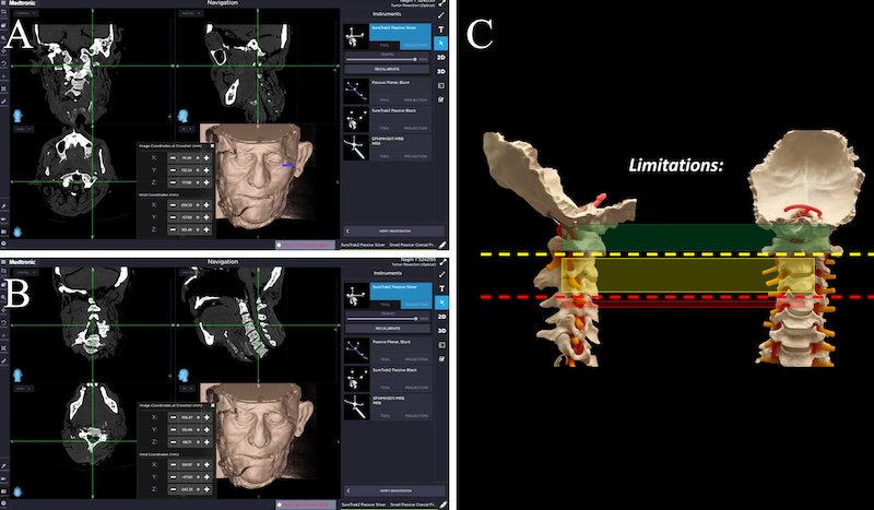
Fig. 2: A navigation snapshot showing the contralateral C1 foramen transversarium line is easily accessible via left ETCOX (A), XYZ values of the most inferior point that can be reached via ETCOX (B), and color coded surgical freedom and limitations (C).
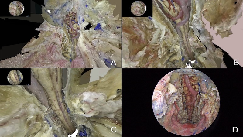
Fig 3: 3D anatomical model created via reality capture showing the ETCOX whole surgical freedom (A,B and C). The 2D endoscopic view via EEA after completing the ETCOX showing the UUCS drilling and upper spinal cord.)
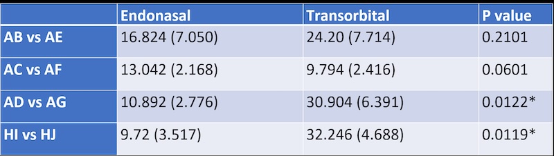
References
Table 1: Mean (standard deviation) were presented.
P values are from Wilcoxon two-sample tests.
*p-value<0.05; **p-value<0.01; ***p-value<0.001
Wilcoxon two-sample tests.
There is no significant difference between the transnasal group vs the transorbital group regarding lateral access (both contralateral and ipsilateral)
There is significant difference between the two groups in terms of caudal access, the transorbital group can reach significantly more caudal on the dorsal aspect of the vertebral body [30.9 (6.39) vs 10.9 (2.78) mm, P=0.01] and on the dura surface [32.2 (4.69) vs 9.7 (3.52) mm, P=0.01]
Naros, Georgios
Meningioma Topography Reveals Grade-Specific Distribution and Recurrence Patterns
Georgios Naros, Sophie Wang, Aldo Spolaore, Kathrin Machetanz, Marcos Tatagiba
Department of Neurosurgery and Neurotechnology, Eberhard Karls University,
Background
High-grade meningiomas (WHO grade II–III) are characterized by a substantially higher risk of tumor recurrence or progression following microsurgical resection compared to low-grade meningiomas. Previous studies have reported differences in their epidemiological and anatomical distribution, including a higher incidence in males and a lower prevalence at the skull base. This study aimed to analyze the spatial distribution of high-grade and recurrent meningiomas using a voxel-based approach.
Methods
This retrospective cohort study included patients with histologically confirmed meningiomas. Preoperative magnetic resonance imaging (MRI) scans were normalized, and tumors were segmented using an automated algorithm. Patient-specific volumes of interest were used to perform voxel-based morphometry (VBM), linking spatial tumor distribution to clinical parameters and outcomes. Diease progression was defined as radiographic tumour progression (in Gd contrast-enhances MRI) Extent of resection was classified according to Simpson grades.
Results
A total of 721 patients were included, of whom 23% had high-grade meningiomas. High-grade meningiomas demonstrated a distinct spatial distribution, with significant voxel clustering in the frontal, temporal, central, and parietal convexity. In contrast, low-grade meningiomas were more frequently located in the skull base, posterior fossa, and occipital convexity. Tumor recurrence or progression was significantly more common in high-grade meningiomas (36% vs. 17%; p<.001), despite a higher extent of resection (in Simpson) in this group, likely reflecting their lower prevalence at the skull base. Recurrence in high-grade meningiomas predominantly occurred in the central and parietal convexity, whereas low-grade meningiomas showed the highest recurrence density in the medial skull base. Subgroup analysis of WHO grade II meningiomas revealed no significant differences in voxel-based spatial distribution between tumors classified as high-grade due to atypia and those classified as high-grade solely on the basis of brain invasion.
Conclusions
High-grade and low-grade meningiomas exhibit distinct anatomical distribution and recurrence patterns on voxel-based analysis. The absence of spatial differences between atypical and brain-invasive WHO grade II meningiomas suggests that brain invasion and atypia may represent related manifestations of aggressive tumor biology, supporting the current WHO classification of brain invasion as an independent criterion for WHO grade II meningioma.
Author Disclosure Information
G. Naros: None. A. Spolaore: None. S. Wang: None. K. Machetanz: None. M. Tatagiba: None.
Nasrallah, Vanessa
Predictive Growth Parameters in Grade 1 Meningioma
Vanessa Nasrallah, Amirhossein Akhavan-Sigari, Alice Giotta Lucifero, Matthew Dufault, Estefana Bcharah, Landon Gray, Mytreyi Trivedi, Bernard Bendok
Mayo Clinic
Introduction
Predicting the growth of WHO grade 1 meningiomas is essential for individualized management, particularly when determining whether active surveillance or early intervention is appropriate. We conducted a scoping review to summarize current evidence regarding imaging and clinical factors associated with tumor growth.
Methods
A literature search of PubMed, Embase, and Cochrane CENTRAL was performed from database inception through 2023. Studies evaluating imaging or clinical predictors of growth in radiologically or histologically confirmed WHO grade 1 meningiomas managed with serial imaging were included. Studies involving higher-grade meningiomas, recurrent tumors, case reports, and non-English publications were excluded. Data regarding study characteristics, patient demographics, imaging biomarkers, growth definitions, and reported predictors were extracted for qualitative synthesis.
Results
A total of 8 studies comprising 1,350 patients met the inclusion criteria. Considerable heterogeneity existed in growth definitions and reporting metrics, precluding quantitative synthesis. Across studies, younger age, larger baseline tumor volume, absence of calcification, T2/FLAIR hyperintensity, lower apparent diffusion coefficient (ADC) values, and selected tumor locations were the most frequently reported factors associated with accelerated tumor growth. Several multivariable prediction models, including the IMPACT model, demonstrated potential for individualized risk stratification but have undergone limited external validation.
Conclusion
Age, location, initial volume, T2/FLAIR intensity, calcification, and ADC are most reliable factors predicting growth of Grade 1 meningiomas. IMPACT model is useful clinically for patient-specific surveillance despite requiring validation and standardization of growth rates.
Orlov, Egor
Intraluminal Endoscopic Anatomy of the Cranial Venous System and Venous Bypass Procedures: An Anatomical Study
Egor Orlov, Natalia Polunina, Michael Fritsch
Dietrich-Bonhoeffer-Klinikum
Introduction/Background
The cerebral venous system is highly variable. Convexity meningiomas more often show higher WHO grades than skull base lesions, making radical resection important for durable tumor control; however, their relationship to bridging veins, venous lacunae, and dural sinuses may limit safe resection. The intraluminal endoscopic anatomy of cranial venous structures remains insufficiently studied.
Objectives
To explore the intraluminal endoscopic anatomy of the cranial venous system in latex- injected human cadaveric heads, identify features relevant to tumor–vein relationships and venous preservation, and assess the anatomical feasibility of selected venous bypass procedures for complex sinus-involving lesions.
Methods/Description
Twenty human cadaveric heads were latex-injected and examined microsurgically and endoscopically. A parasagittal craniotomy was performed along the superior sagittal sinus, followed by dissection of first- and second-order venous tributaries. Venous entry sites, intertributary distances, and insertion angles were measured. All dural sinuses were explored intraluminally with video-recorded endoscopy and analyzed for ostia, septations, trabeculations, morphology, and variability.
Results/Comments
Endoscopy demonstrated marked variability of the superior sagittal sinus. We propose five descriptive patterns: simple tubular, trabecular, septated, lacunar-compartmental, and complex mixed. The simple tubular pattern was most frequent (60%). In 20%, the anterior third showed pronounced septation with intraluminal sinus duplication.
Conclusion
Intraluminal endoscopy identifies high-risk sinus segments characterized by septations, trabeculations, or duplication. These regions may be surgically vulnerable because complex intraluminal anatomy could promote local flow slowing and postoperative thrombosis after sinus manipulation. This insight may support safer resection, venous preservation, and selected venous bypass planning in sinus-involving meningiomas.
Pandit, Anand
Stereotactic Radiosurgery for Paediatric Meningiomas: A Systematic Review of Safety and Efficacy
Anand Pandit, Holly Aylmore, Jia Nag, Magda Segura Arderiu, Matthew Short, Patrick Grover
National Hospital for Neurology and Neurosurgery
Background
Paediatric meningiomas are rare, accounting for 1-5% of all meningiomas and 1-2% of paediatric brain tumours. While surgical resection remains the primary treatment, gross total resection is not always achievable and the role of adjuvant stereotactic radiosurgery (SRS) in children is poorly defined. This systematic review synthesises available evidence on the safety and efficacy of SRS in paediatric meningiomas to inform clinical decision making and identify research priorities.
Methods
A systematic search of MEDLINE, Embase, Scopus, and Web of Science was conducted from inception to March 2025, following PRISMA guidelines (PROSPERO: CRD420251030411). Original research studies and case series reporting SRS in patients under 19 years old with meningiomas were included. Risk of bias was assessed using the MINORS tool. Findings were synthesised narratively owing to study heterogeneity.
Results
Eight retrospective studies encompassing 100 patients (age range 2-19 years) were included. All studies were rated moderate risk of bias. Meningiomas were reported across a broad range of intracranial locations with volumes between 0.6-25.5cm3. SRS was predominantly delivered as adjuvant therapy following subtotal resection. The largest study (n = 57) reported local tumour control in 85.9% of cases, with 5 year progression-free survival (PFS) of 86% and 10 year PFS of 69%. Ten year overall survival was 92.1%. Adverse radiation effects were reported in 8.8% of patients in the one study that formally assessed them. Substantial heterogeneity in SRS platform, dose (10-30Gy), follow up reporting, and outcome definitions precluded meta-analysis.
Conclusion
SRS appears feasible and associated with meaningful tumour control in selected paediatric meningioma patients, with outcomes broadly comparable to adult series. However, the evidence base is limited by small retrospective cohorts, inconsistent outcome reporting, and a near-total absence of long term safety data. Prospective, standardised, multicentre studies are needed to establish best practice in this population.
Prabhu, Vikram
Pre-Operative Embolization of Meningiomas: Single-Center 10-Year Experience
Ronak Jani, John Tsiang, Kenneth Jordan, Aalim Khaderi, Danielle Baffa, Brandon Bond, Anthony Kam, Joseph Serrone, Min Park, Vikram Prabhu
Loyola University Medical Center
Abstract
Background
Large or anatomically complex meningiomas may pose considerable surgical challenge with risk for high intraoperative blood loss. Pre-operative angiography and endovascular embolization may aid resection by reducing tumoral bleeding, inducing necrosis, and detailing surrounding neurovasculature.
Objectives
We sought to describe our 10-year experience with selectively performing pre-operative endovascular embolization for meningiomas at a single academic, tertiary care center.
Methods
Chart and imaging review was conducted for all consecutive patients with available records who underwent pre-operative endovascular embolization followed by surgical resection for pathologically confirmed meningioma at our center from 2016 to 2026. Demographics, tumor characteristics, embolization data, surgical data and outcomes, and hospitalization outcomes were collected. WHO grade 1 vs. grade 2 cases were compared.
Results
We analyzed 28 patients. Mean tumor volume was 65.9 mL. 46.4% were skull base meningiomas. 35.7% were WHO grade 2. Embolization achieved mean 71.3% devascularization with 50% achieving ≥ 90% devascularization. Surgical resection rates were excellent, with mean volumetric resection 98.5%. Gross total resection was achieved in 64.3%, and 57.1% were Simpson grade 1 resections. Mean estimated blood loss was 245.5 mL; only 10.7% required transfusion. Mean surgery duration was 349.0 minutes. Hospitalization lasted mean 12.3 days and 50% were discharged home. Zero complications from embolization occurred. There were no statistically significant differences in outcomes for WHO grade 1 vs. WHO grade 2 patients.
Conclusion
Pre-operative embolization can be safely and effectively performed for selected meningiomas with excellent endovascular and surgical outcomes for both WHO grade 1 and WHO grade 2 tumors.
Rahmanov, Serdar
Lingual Neuralgia as an Atypical Manifestation of Trigeminal Neuralgia: Two Cases With Posterior Fossa-to-Tongue V3 Anatomical Correlation
Serdar Rahmanov, Evgeniia Rahmanova, Safwan Alomari, Badih Adada, Hamid Borghei-Razavi
Cleveland Clinic Florida
Background
Trigeminal neuralgia classically causes unilateral electric shock-like facial pain in one or more trigeminal divisions. Isolated tongue-predominant pain is rare and may be mistaken for dental, oral, otolaryngologic, glossopharyngeal, or burning mouth disorders. The lingual nerve, a branch of the mandibular division (V3), carries sensation from the anterior two-thirds of the tongue, providing a pathway for lingual neuralgia.
Objectives
To report two cases of tongue-predominant trigeminal neuralgia and correlate the presentation with the V3–lingual nerve pathway using cadaveric illustrations.
Methods
Two patients with unilateral tongue-predominant neuralgic pain were reviewed. Clinical features, triggers, interventions, imaging, operative findings, and outcomes were analyzed. Cadaveric dissections of V3 and the lingual nerve illustrated continuity between the trigeminal root, mandibular division, lingual nerve, and anterior tongue territory.
Results
A 63-year-old man with longstanding trigeminal neuralgia developed left tongue pain with constant neuropathic discomfort and electric shock-like attacks triggered by speech, eating, and thermal stimuli. MRI showed neurovascular contact at the trigeminal root entry zone without marked deformity. Further radiosurgery was discouraged because of concern for worsening neuropathic pain. A 48-year-old man with multiple sclerosis and prior Gamma Knife radiosurgery developed recurrent paroxysmal left tongue pain triggered by speaking, chewing, swallowing, and tooth brushing. MRI demonstrated compression by superior cerebellar artery branches. Microvascular decompression revealed superior cerebellar and anterior inferior cerebellar artery compression, followed by substantial improvement.
Conclusion
Tongue-predominant trigeminal neuralgia may occur through the V3–lingual nerve sensory pathway. Recognizing this anatomical-clinical relationship may prevent delayed diagnosis and guide treatment. Figure 1
Rakholia, Yogesh
Differential RNA Expression in Meningiomas in Patients With Breast Cancer
Yogesh Rakholia, Hayden M. Dux, Harsh Shah, Selvaraj Muthusamy, Hope Richard, William C. Broaddus
Virginia Commonwealth University
Background
Meningiomas are the most common primary central nervous system tumors in adults, comprising 36% of new diagnoses. Breast cancer is the most common cancer diagnosis in the United States, affecting 13% of American women. There is markedly increased incidence of breast cancer in patients with a history of meningioma, and vice versa, compared to the baseline population, suggesting shared molecular mechanisms. Objective: Compare single-cell RNA transcriptomes between patients with meningioma with or without concomitant breast cancer to identify possible cellular pathways explaining the increased susceptibility of patients with breast cancer to develop meningioma.
Methods
Endogenous mRNA sequencing using a NanoString (Seattle, WA) targeted gene expression panel was performed on surgical pathology samples obtained from meningioma and breast tumor resections. Gene enrichment was analyzed using Ingenuity Pathway Analysis in QIAGEN.
Results
There were 22 differential expressed genes (DEGs) between meningioma samples from patients with and without breast cancer: 18 genes were upregulated and 4 were downregulated (p<0.05, log2 (fold change) >0.5 or < -0.5). The most significant upregulated gene was BAD and the most significant downregulated gene was MAPK14. 28% (5/18) of upregulated DEGs (APC, FASLG, SHC1, IGF1R, RPS6KA1) in this cohort are known to be enriched in breast cancer samples (p = 0.0032). The top canonical pathways include PI3K/AKT signaling, PTEN signaling, and HER-2 signaling (p<0.05).
Conclusion
Differential gene expression study provides insight into genetic interplay between meningiomas and breast cancer to potentially reveal novel biomarkers for early detection, prevention, and therapeutic targets.
Sabbagh, Aria
Histotripsy for Tumor Ablation and Enhancement of Immunotherapy
Aria Sabbagh, Prapannajeet Biswal, Tarana Kaovasia, Prudhvi Mallepaddi, Sai Samala, Khadijeh Koushki, Geraldine Raja, Nitin Tandon, Zhen Xu, Sunil Krishnan
UTHealth Houston
Introduction
Glioblastoma remains fatal, and immune checkpoint inhibitors have failed to provide meaningful survival benefit, largely due to the tumor’s immunosuppressive microenvironment. Histotripsy, a non-thermal focused ultrasound technology, induces mechanical tumor fragmentation, opens the blood-brain barrier, and preserves antigen integrity. Histotripsy is FDA-approved for treatment of liver tumors, achieving a primary efficacy of 95.5% in clinical trials (PMID 39225612).
Objective
To evaluate the efficacy of histotripsy for brain tumor ablation and determine whether its combination with immunotherapy enhances anti-tumor immune responses and improves survival.
Methods
C57BL/6 mice underwent stereotactic intracranial implantation of GL261 glioma cells, followed by treatment with saline, histotripsy, anti-PD-1, or the combination of histotripsy and anti-PD-1. Tumor growth was monitored using in vivo bioluminescence imaging, and mice were monitored for survival. Immune responses were assessed by flow cytometry of brain, spleen, blood, and bone marrow samples.
Results
Combination of histotripsy and anti-PD-1 significantly reduced tumor growth compared with PBS and monotherapy groups (p<0.05, one-way ANOVA), and this was associated with a significant improvement in murine survival (p<0.05, Mantel-Cox Test). Immune profiling revealed an increase in activated T cells and a decrease in exhausted T cells in the brain and peripheral immune compartments (p<0.05, one-way ANOVA). The tumor associated myeloid compartment was reprogrammed toward a more pro-inflammatory, less immunosuppressive phenotype (p<0.05).
Conclusions
Histotripsy synergizes with immunotherapy to enhance anti-tumor immunity, suppress glioma progression, and improve survival. These findings provide both mechanistic and translational rationale for a new class of CNS-directed immune priming strategies, offering hope to patients for whom no effective immunotherapy exists.
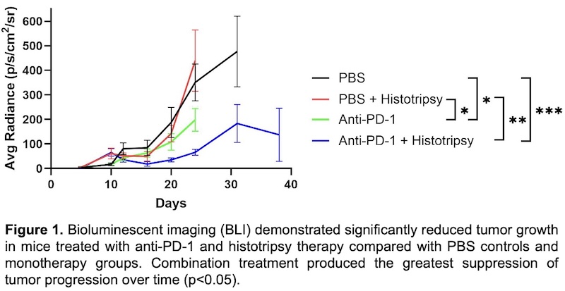
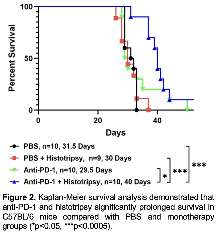
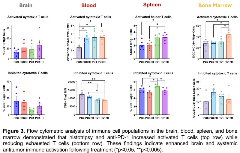
Sahin, Mustafa Selim
A Low-Cost Sheep Brain Model for Acquisition of Fundamental White Matter Dissection Skills: A Prospective Educational Pilot Study
Mustafa Selim Sahin, Latif Saglam, Deniz Akpınar, Fatih Koksoy, Cafer Ikbal Gulsever, Duran Sahin, Duygu Dolen Burak, Osman Coskun, Ozcan Gayretli, Tugrul Cem Unal
Istanbul University Istanbul Faculty of Medicine
Introduction
White matter dissection is a cornerstone of microsurgical neuroanatomy education. However, access to cadaveric specimens is limited and novice trainees often lack basic dissection skills before entering cadaver laboratories. We evaluated a low-cost sheep brain model prepared with a modified Klingler technique as a preliminary training platform for white matter dissection.
Objectives
To assess whether repeated sheep brain dissection sessions improve fundamental white matter dissection skills in participants without prior white matter dissection experience.
Methods
Ten participants underwent three standardized dissection sessions at one-week intervals. During each session, cortical decortication and U-fiber dissection were performed within a predefined 3×3 cm frontal/temporal region. Performance was evaluated using dissection time and preservation of the white matter dissection plane. Plane violation was defined as continuation of dissection despite a visible change in fiber orientation following U- fiber removal.
Results
Mean dissection time(minute) decreased from 42.2 during the first session to 35.9 during the second session and 27.1 during the third session. Preservation of the correct dissection plane improved throughout training, increasing from 32% in the first session to 65% in the second session and 100% in the final session.
Conclusion
Repeated practice on sheep brains prepared using a modified Klingler technique resulted in substantial improvements in dissection efficiency and preservation of the appropriate white matter plane. This inexpensive and reproducible model may serve as an effective preparatory step before cadaveric white matter dissection training.
Shigeeda, Ryota
Usefulness of combining the transnasal-transsphenoidal and supraorbital approaches for resection of tuberculum sella meningiomas
Ryota Shigeeda, Takuichiro Hide
Department of Neurosurgery, Indiana University School of Medicine
Background
The surgical approach to treating tuberculum sella meningiomas must be determined based on the extent to which they extend into the sella, laterally and anteriorly. The transnasal-transsphenoidal approach is effective for tumors extending into the sella or posteriorly, while the transcranial approach is effective for tumors extending laterally or anteriorly.
Objectives
To reap the benefits of both approaches, we performed surgery using a combination of a transnasal-transsphenoidal approach and a supraorbital approach.
Methods
The patient was a 49-year-old woman with a tuberculum sella meningioma that had extended from the intrasellar region over the lateral aspect of the right internal carotid artery. We began the procedure transnasally, first detaching the tumor from the tuberculum sella in order to cut off its blood supply. We then dissected the tumor simultaneously using both the transnasal and supraorbital approaches. This allowed us to confirm each other’s progress. Finally, we removed the tumor as a single mass via the transnasal corridor.
Comments
Attempting to treat a case like this using the transnasal approach alone would require an extended transnasal-transsphenoidal technique, thereby increasing the risk of complications, including cerebrospinal fluid leaks. Furthermore, craniotomy alone does not provide sufficient visualisation of the intrasellar region. Although having two surgeons present is inconvenient, performing both the transnasal and transcranial procedures simultaneously enabled us to perform minimally invasive surgery.
Conclusion
Performing both the transnasal-transsphenoidal and the supraorbital-subfrontal approaches simultaneously would enable us to manage tuberculum sella meningiomas regardless of their direction of extension.
Sinha, Mousumi
Predictors of Early Neurosurgical Escalation in Idiopathic Intracranial Hypertension: A Retrospective Cohort Analysis of Patients from the EPIC COSMOS Data Model
Mousumi Sinha, Rahil Tai
The University of Texas Health Science Center at Houston
INTRODUCTION / BACKGROUND
Idiopathic intracranial hypertension (IIH) is a disorder of elevated intracranial pressure without structural cause, predominantly affecting young obese women. Population-level predictors of early surgical escalation remain poorly defined across procedure categories.
OBJECTIVES
To identify independent clinical, laboratory, and utilization predictors of early neurosurgical escalation within 90 days of IIH diagnosis.
METHODS / DESCRIPTION
Retrospective cohort study using EPIC COSMOS (2018–2025); adults with IIH (ICD-10 G93.2) and ≥2 specialty encounters in Neurology, Ophthalmology, or Neurosurgery. Primary outcome: surgical escalation within 90 days of IIH index date (CPT codes: VP shunt 62223/62230, LP shunt 63741, VSS 61635/37252/37253, ONSF 67300/67570). Secondary outcomes included time-to-surgery refractory categories and post-surgical morbidity. Multivariable logistic regression with multiple imputation for missing BMI (14%; MICE, m=5). Sensitivity analysis excluded concurrent pregnancy.
RESULTS / COMMENTS
Of 190,236 patients (mean age 40, 86.4% female, mean BMI 36.8, obesity 73.1%), 3,694 (1.9%) underwent surgical escalation within 90 days (sensitivity cohort 2.0%, identical). Multivariable logistic regression (N=186,912, AUC=0.754) identified the following independent predictors:
- Strongest predictors: cerebral venography OR 4.38 (95% CI 3.61-5.30), therapeutic LP OR 3.24 (2.86-3.67), hypokalemia OR 2.53 (2.33-2.76), anemia OR 2.28 (2.11-2.46), acetazolamide use OR 1.82 (1.69-1.96) — all p<0.001
- Protective factors: migraine OR 0.77 (0.72-0.83, p<0.001) and higher Social Vulnerability Index OR 0.84 (0.75-0.95, p=0.005), indicating a health disparity in surgical access
- Of 9,840 ever-surgical patients, 37% (n=3694) had surgical escalation within 90days of IIH index date. VSS was the dominant procedure in this group (n=1,706, 47.7%)
Post-surgical morbidity was substantial: 30-day readmission 7.7%, ED utilization 66%, ICU admission 32%. Mean LOS ranged from 5.3 days (VSS) to 6.8 days (ONSF)
CONCLUSION
In the largest real-world IIH cohort, cerebral venography, therapeutic LP, and electrolyte disturbances independently predicted early neurosurgical escalation. Over one-third of surgical patients had chronic/delayed escalation (mean 3.2 years), revealing a critical referral gap and health equity disparity in surgical access.
Sousa, Sergio
From Repurposed Microscopes to a Microsurgical and Skull Base Training Laboratory: A Sustainable Model for Neurosurgical Education
Sergio Sousa, Joao Silva, Vasco Pinto, Alfredo Calheiros
Hospital de Santo António
Introduction
Microsurgical and skull base training are essential components of neurosurgical education, yet dedicated laboratories often require substantial financial and infrastructural resources. We describe the development of a neurosurgical training laboratory built from the ground up through resource reutilization, progressive institutional support, and academic collaboration.
Methods
In 2021, our department had no dedicated microsurgical laboratory or specific funding. The project began by recovering a discontinued operating microscope and repurposing obsolete hospital materials for simulation-based training. A second discontinued microscope was subsequently recovered, expanding training capacity. Educational activities were progressively developed alongside the laboratory.
Results
The initial single-station, resource-limited initiative evolved into a structured microsurgical training platform. Since 2025, the laboratory has hosted an annual microneurosurgical course focused on microvascular suturing and anastomosis, with its third edition planned for February 2027. Following the success of the initiative, funding was secured for three new microscopes, all of which were incorporated into the most recent course edition, significantly expanding training capacity. Most recently, a partnership with the Abel Salazar Institute of Biomedical Sciences (ICBAS) enabled the launch of an annual cadaveric skull base training course for our institution’s neurosurgical residents. The inaugural edition focused on temporal bone approaches, particularly anterior and posterior petrosectomies, further expanding the laboratory’s role in hands-on anatomical and surgical training.
Conclusion
Our experience demonstrates that limited initial resources need not preclude the development of a high-quality neurosurgical training environment. Resource reutilization, incremental growth, institutional support, and academic partnerships transformed an initially low-cost initiative into an expanding platform integrating microvascular and cadaveric skull base training.
Sulaimanov, Umid
Complementary Corridors to the Sellar Region: A Cadaveric Comparison of Fronto-Orbital and Pterional Transsylvian Approaches
Umid Sulaimanov, Mariagrazia Nizzola, Nafiye Sanlier, Nicola Boari, Pietro Mortini, Abdullah Keles, Ufuk Erginoglu, Mustafa K Baskaya
Department of Neurological Surgery, School of Medicine and Public Health, University of Wisconsin-Madison
Introduction/Background
Transcranial approaches to the sellar and parasellar region remain essential for selected lesions requiring optic pathway decompression, vascular control, or access to retrosellar extensions. The fronto-orbital (FO) and pterional transsylvian (PT) routes offer distinct operative corridors with different anatomical advantages.
Objectives
To compare FO and PT corridors for sellar and parasellar exposure through cadaveric morphometric and corridor-based anatomical analysis.
Methods/Description
Twelve formalin-fixed, silicone-injected cadaveric specimens (24 sides) underwent bilateral FO and PT approaches. Microsurgical dissections were performed under operative microscopy, with corridors documented stepwise. Operative depth to predefined sellar, optic pathway, and retrosellar landmarks was measured using digital calipers and compared with paired two-tailed t-tests. Qualitative exposure was independently rated by two observers.
Results/Comments
Both approaches reliably exposed the sellar region, with comparable depth to the diaphragma sellae opening (FO 49.5±2.26 mm vs PT 50.1±2.60 mm). FO provided shorter access to anterior and contralateral optic pathway structures, including the contralateral optic canal (42.9±2.29 mm vs 48.8±1.65 mm). Conversely, PT afforded shorter working distances to posterior sellar and retrosellar structures, including the posterior clinoid process (46.9±1.89 mm vs 57.8±2.21 mm). FO primarily exploited the prechiasmatic route, whereas PT provided broader access through the opticocarotid and carotid-oculomotor corridors.
Conclusion
FO and PT approaches provide complementary rather than interchangeable access to the sellar and parasellar region. FO favors bilateral optic pathway and anterior midline exposure, whereas PT facilitates retrosellar and vascular access, supporting individualized corridor selection based on lesion anatomy and operative goals.
Keywords
Sellar region; parasellar region; surgical corridor; skull base anatomy; fronto-orbital approach; pterional transsylvian approach
Sulaimanov, Umid
External Validation of the Foramen Magnum Meningioma Risk Score (FMMRS): Good Ranking but Poor Calibration
Umid Sulaimanov, Nafiye Sanlier, Behman Demir, Ahmed Rasim Bayramoglu, Abdullah Keles, Ufuk Erginoglu, Mustafa K Baskaya
Department of Neurological Surgery, University of Wisconsin-Madison
Background
Foramen magnum meningiomas (FMMs) are histologically benign but carry substantial perioperative morbidity owing to their intimate neurovascular relationships. The Foramen Magnum Meningioma Risk Score (FMMRS) is the only published preoperative, MRI-based score for estimating postoperative complication risk, yet its original validation was small, not fully independent, and assessed discrimination without calibration.
Objectives
To perform a fully independent external validation of the FMMRS, adding a calibration assessment of its published absolute-risk estimates.
Methods
In this single-center retrospective study, 40 consecutive adults who underwent microsurgical FMM resection were analyzed. The FMMRS was recalculated de novo from preoperative MRI. Two endpoints were prespecified: any complication and major complication. Discrimination was assessed for both endpoints using the area under the ROC curve (AUC). Calibration was assessed for the any-complication endpoint, for which the published estimates were derived. Threshold performance and the Cochran-Armitage trend test were also evaluated.
Results
Any complication occurred in 14/40 patients (35.0%) and major complication in 7/40 (17.5%). Discrimination was modest (AUC 0.64 and 0.67). Calibration was poor, with systematic overestimation of absolute risk; the largest discrepancy was in the high-risk group, where expected and observed rates were 96.4% versus 42.9%. In exploratory analysis, FMMRS ≥6 captured all major complications (sensitivity and negative predictive value both 100%), whereas the published threshold of ≥8 missed nearly half.
Conclusion
The FMMRS retained only limited value as an ordinal stratifier and substantially overestimated absolute complication probabilities. Its published probabilities should not guide individualized preoperative counseling without recalibration; larger multicenter, patient-level studies are required.
Sulaimanov, Umid
Perfusion Before Resection: A Flow-Guided Microsurgical Strategy for Intracranial Infectious Aneurysms
Umid Sulaimanov, Mariagrazia Nizzola, Nafiye Sanlier, Yerkebulan Serikkanov, Ufuk Erginoglu, Abdullah Keles, Mustafa K Baskaya
Department of Neurological Surgery, University of Wisconsin-Madison
Background
Intracranial infectious aneurysms (IIAs) pose a unique operative challenge because the diseased segment is often fragile, inflamed, distally located, and involves small-caliber cortical branches. The central question is not only how to eliminate the aneurysm, but whether distal perfusion can be maintained after trapping and excision.
Objectives
To describe a flow-guided operative strategy for IIAs and show how intraoperative assessment guides the choice between bypass reconstruction and excision without revascularization.
Methods
An institutional retrospective review was performed of patients treated microsurgically for IIAs from January 2006 to November 2025. Clinical presentation, aneurysm location, preoperative imaging, intraoperative flow-assessment methods, reconstructive strategy, postoperative angiography, and neurological outcomes were reviewed.
Results
Three patients with distal middle cerebral artery infectious aneurysms were included. Two presented with ruptured aneurysms causing intraparenchymal and subarachnoid hemorrhage; one had an enlarging unruptured aneurysm with infective endocarditis. All aneurysms were exposed, trapped, and excised. Intraoperative decision-making used indocyanine green videoangiography, micro-Doppler ultrasonography, temporary occlusion, neurophysiological monitoring, and, in selected cases, a surrogate balloon test occlusion with hypotensive challenge. Flow replacement was required in two patients: one underwent intracranial-intracranial bypass with an interposition superficial temporal artery graft, another in situ reconstruction with distal and proximal anastomoses. In the third, preserved collateral perfusion during prolonged temporary occlusion allowed safe excision without bypass. Postoperative angiography confirmed obliteration with preserved distal perfusion or bypass patency.
Conclusion
Microsurgical treatment of IIAs should be guided by real-time collateral-flow tolerance rather than morphology alone, supporting selection among bypass, in situ reconstruction, or excision.
Tawk, Rabih
Monitored Outpatient Recovery After Elective Neuroendovascular Procedures: Safety and Cost Analysis
Rabih Tawk, Rafael Tatit, Ahmad Khatib, Baher Boktor, Anshum Patel, Hani Alhaj-Zain, Jackie Dean, Monish Vijayaraghavan, William D. Freeman
Mayo Clinic
Introduction
Routine overnight admission after elective neuroendovascular procedures consumes acute-care capacity. We compared monitored outpatient recovery in an on-campus Care Hotel versus inpatient observation for safety, acute-care utilization, and modeled facility costs.
Methods
We studied adults undergoing selected elective neuroendovascular procedures from August 2019 through August 2025. The Care Hotel cohort comprised 100 consecutive pathway- eligible patients transferred after 4 hours of postprocedural observation. Controls were retrospectively selected 1:1 from otherwise eligible patients who underwent inpatient observation before pathway implementation or for logistical reasons (54 historical, 46 contemporaneous). The primary endpoint was 30-day major neurologic adverse event. Facility costs were modeled from interventional-suite and post-PACU recovery-destination times using external unit costs.
Results
Procedure categories were balanced between Care Hotel and inpatient-observation groups (maximum absolute SMD, 0.154); aneurysm treatment was most common (67.0% and 74.0%). The primary endpoint occurred in 2.0% vs 6.0% (adjusted OR, 0.40; 95% CI, 0.09–1.87; P=.230). Thirty-day complications occurred in 6.0% vs 8.0%; ED visits, 14.0% vs 13.0%; and procedure-related readmissions, 3.0% vs 3.0%. Median hospital time from admission to discharge was 10.0 vs 28.8 hours (P<.001). Mean modeled facility cost per episode of care was $9,694 vs $14,249, and $10,045 vs $14,633 including 30-day costs (both P<.001).
Conclusion
Monitored outpatient recovery was not associated with higher 30-day rates of adverse events or acute-care utilization, and was associated with shorter hospital time and lower modeled facility costs. These findings support prospective validation.
Turel, Keki
Anatomy of the Tentorium: Classification and Strategy for Surgical Approaches in the Management of Tentorial Meningiomas
Keki Turel
BOMBAY HOSPITAL
Introduction/Background
Tentorial meningiomas constitute a small proportion of intracranial meningiomas but remain among the most challenging skull base tumors because of their close relationship with the brainstem, cranial nerves, deep venous system, and major dural venous sinuses. Successful surgery requires a detailed understanding of tentorial microsurgical anatomy and meticulous selection of the operative approach.
Objectives
To present a practical anatomical classification of tentorial meningiomas and describe an individualized strategy for selecting the optimal surgical approach based on tumor location, extent, neurovascular relationships, and vascular supply.
Methods/Description
Based on Prof. Dr. Keki E. Turel’s extensive microsurgical experience, the tentorium is analyzed anatomically, and tentorial meningiomas are classified according to their dural attachment, anatomical compartment, and predominant arterial blood supply. Surgical approaches—including supracerebellar infratentorial, occipital transtentorial, subtemporal, retrosigmoid, and combined skull base techniques—are correlated with tumor location. Emphasis is placed on early identification and control of feeding arteries, preservation of venous sinuses, cranial nerves, arterial perforators, and the brainstem to achieve maximal safe resection.
Results/Comments
The proposed anatomy-based classification facilitates individualized surgical planning, optimal operative exposure, safer microsurgical dissection, and selection of the most appropriate surgical corridor. This strategy minimizes neurological morbidity while maximizing safe tumor removal and preserving critical neurovascular structures.
Conclusion
A comprehensive understanding of tentorial anatomy, tumor classification, and vascular anatomy provides a reliable framework for individualized surgical management of tentorial meningiomas, improving surgical safety, extent of resection, and overall patient outcomes.
Turel, Keki
Parasagittal Meningiomas
Keki Turel
BOMBAY HOSPITAL
Introduction
Parasagittal meningiomas constitute nearly one-quarter of intracranial meningiomas and remain among the most challenging lesions in cranial neurosurgery. Their intimate relationship with the superior sagittal sinus (SSS), cortical draining veins, and eloquent cortex creates a delicate balance between maximal tumor removal and preservation of venous circulation and neurological function.
Objective
To review the surgical anatomy, classification, operative nuances, and individualized management of parasagittal meningiomas according to the location and extent of SSS involvement.
Methods
Based on Prof. Dr. Keki E. Turel’s extensive surgical experience, this presentation emphasizes detailed preoperative assessment using MRI and MR venography to determine sinus patency, collateral venous drainage, tumor location, and cortical vein anatomy. Microsurgical dissection, neuronavigation, careful preservation of draining veins, and tailored strategies for tumors involving the anterior, middle, or posterior third of the SSS are discussed.
Results
Favorable outcomes depend on individualized planning. Gross total excision may be achieved when sinus invasion is limited and venous drainage can be safely preserved. In extensive sinus involvement, subtotal resection followed by stereotactic radiosurgery may provide durable tumor control. However, response to radiosurgery, including proton beam therapy, is variable and influenced by tumor biology. Histopathological and molecular analysis therefore remain essential for prognosis, adjuvant treatment selection, and long-term management. Preservation of venous drainage is the principal determinant of neurological outcome and surgical morbidity.
Conclusion
Parasagittal meningiomas remain an enigma because no single strategy suits every patient. Success requires precise anatomical knowledge, meticulous microsurgical technique, rational patient selection, tumor biopsy, and judicious multimodal therapy.
Yamamoto, Junichi
Extradural Anterior Clinoidectomy and Optic Canal Decompression for Clinoidal Meningioma with Optic Canal Invasion: A Surgical Video
Junichi Yamamoto
Albany Medical College/Albany Medical Center
Introduction/Background
Clinoidal meningiomas may cause visual symptoms when they extend into the optic canal and compress the optic nerve. We present a surgical video of a right clinoidal meningioma in a woman in her 40s who presented with a right temporal visual field defect. The tumor measured 3.4 × 2.9 × 3.0 cm and demonstrated optic canal invasion with clinically significant optic nerve compression.
Objectives
To demonstrate, through a surgical video presentation, the usefulness of extradural anterior clinoidectomy and complete optic canal decompression for safe tumor removal and optic nerve decompression in a symptomatic clinoidal meningioma with optic canal invasion.
Methods/Description
The surgical video demonstrates early extradural anterior clinoidectomy performed before intradural tumor manipulation. This approach allowed early devascularization of the tumor by interrupting its dural blood supply around the anterior clinoid process and optic canal. It also provided safe early exposure and proximal control of the internal carotid artery. The optic canal was decompressed from proximal to distal, including the falciform ligament region. After complete bony decompression, the optic sheath was opened to further decompress the intracanalicular tumor component.
Results/Comments
To minimize thermal injury to the optic nerve, drilling was kept to a minimum. Whenever possible, the anterior clinoid process and optic canal roof were removed using rongeurs after adequate thinning and mobilization of the bone. This drill-sparing, rongeur-based technique facilitated safe optic nerve decompression before tumor manipulation.
Conclusion
This surgical video highlights extradural anterior clinoidectomy with complete optic canal decompression as a useful strategy for clinoidal meningiomas with optic canal invasion. In selected cases, minimizing drilling and using rongeurs may reduce thermal risk to the optic nerve while enabling effective decompression and safe tumor removal.