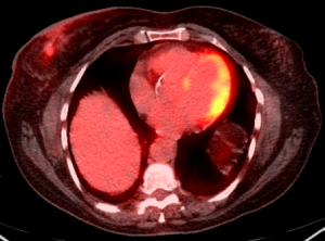
PET in Breast Cancer Detection
Breast cancer forms in tissues of the breast—usually in the ducts, tubes that carry milk to the nipple, and lobules, the glands that make milk. It occurs in both men and women, although male breast cancer is rare. According to estimates by the National Breast Cancer Foundation, Inc., nearly 246,660 people in the United States were diagnosed with breast cancer and more than 40,000 people died from the disease.
Over 2.8 million breast cancer survivors are alive in the United States today. For more facts about breast cancer click here, National Breast Cancer Foundation, Inc.
How is PET used for breast cancer?
Physicians use PET and PET-CT studies to:
- Diagnose and stage of breast cancer by determining the exact location of a tumor, the extent of the disease and whether the cancer has spread in the body, especially the lymph nodes.
- Plan treatment: by selecting the most effective therapy based on the unique molecular properties of the disease and of the patient’s genetic makeup and to determine a site that is appropriate for biopsy, if necessary.
- Evaluate the effectiveness of treatment: by determining the patient’s response to specific drugs and ongoing therapy. Based on changes in cellular activity observed on PET-CT images, treatment plans can be quickly altered.
- Manage ongoing care: by detecting the recurrence of cancer if the patient has new symptoms or shows new abnormalities on other imaging modalities like Ultrasound, CT scan or MRI.
What are the advantages of PET studies for breast cancer Patients?
PET helps physicians and their patients:
- Gain a clear understanding of where and how aggressive the disease is.
- Select a course of treatment.
- Determine the effectiveness of treatments after one or more cycle treatment.
Eliminate unnecessary surgeries after treatment by distinguishing active tumors from residual indicative breast tissue changes.
For more details on how PET is used for breast cancer, visit the Society of Nuclear Medicine and Molecular Imaging.
