Viability Dyes
Unless you are specifically studying apoptosis and cell death, you should include only healthy viable cells in your flow analysis and cell sorting experiments (Fig.1.) because dead and dying cells often show:
- Increased nonspecific antibody binding
- Increased autofluorescence
- Altered phenotype/epitopes
- Compromised RNA/DNA quality and proliferation capacity in downstream applications
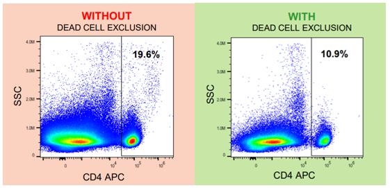
Fig. 1. Artificially high CD4 counts due to nonspecific antibody binding to dead cells. Accurate flow analysis requires exclusion of dead cells.
Relying solely on forward and side scatter to identify healthy cells is not very accurate (Fig. 2.). Further, there are a wide variety of commercially available viability dyes to choose from so it is easy to fit them even into complex multicolor panels.
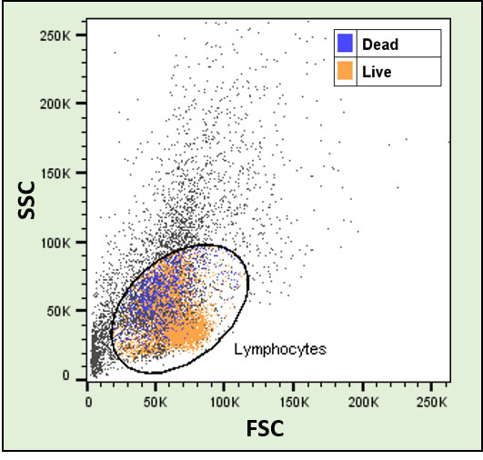
Fig. 2. Cells were isolated from mouse spleen and stained for multiple lymphocyte markers as well as for viability. Live and dead (CD45+ CD3+) T cells could not be separated based on scatter alone.
The specificity of most common viability dyes is based on the integrity of the cell membranes. Healthy cells exclude the dyes and stain only weakly if at all, whereas dying or dead cells have leaky membranes and the dye will penetrate into the cells giving rise to high intensity staining. Note that cell debris may remain unstained and therefore it may sometimes be useful to stain (also) the live cells. It is important to titrate your viability dyes, just like your antibodies.
Viability dyes come in three flavors:
- Cell impermeant DNA binding dyes – stains dead cells (simple to use but cannot be combined with fixation or intracellular staining)
- Cell permeant enzyme substrates – stains live cells (simple to use but cannot be combined with fixation or intracellular staining)
- Cell impermeant amine reactive dyes – stains dead cells (may require extra steps in the staining protocol but can be combined with intracellular staining and fix/perm protocols)
Additional Information:
- Thermo Fisher: BestProtocols: Viability staining protocol for flow cytometry (external link)
- Thermo Fisher: Cell viability assays for flow cytometry (external link)