
Balloon Eustachian Tuboplasty
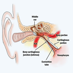
The Eustachian tube (ET) is a narrow tube that connects middle ear space, located behind the tympanic membrane (or ear drum) to the nasopharynx (the space at the back of the nasal passages. This tube serves to equalize the pressure in the middle ear space with external atmospheric pressure. In addition, the tube drains mucus and secretions from the middle ear space.
Mostly, the ET remains closed. It opens when a person swallows, chews or yawns. As it opens, the pressure equalizes and mucus may drain.
When ET cannot perform these functions well, the condition of Eustachian tube dysfunction (ETD) results. Patients with ETD describe ear fullness, hearing loss, tinnitus (ringing sensation) and even dizziness. Many patients with ETD report that they feel as if they are “under water,” and that they cannot clear that their ears.
Not all patients with these symptoms have obstructive ETD. Thus, a good ear exam (known as otoscopy) and special test (tympanometry) are necessary to confirm the diagnosis.
If patient has confirmed ETD, the he or she may be a candidate for a procedure designed to alleviate the symptoms. Traditionally, a myringotomy (a cut in the ear drum), mostly commonly with placement of a pressure-equalizing tube (known as a “PE tube”), has been the primary treatment. This procedure may be performed under local anesthesia in the office setting. Most PE tubes are designed to spontaneously dislodge over the course of a few months or a year. If the PE tube comes out too soon, the ETD may return. Usually, the ear drum will heal, but on occasion, that healing may leave a permanent hole (known as a “tympanic membrane perforation.)
Recently a new procedure, balloon eustachian tuboplasty, has been introduced. In this procedure, a balloon device is introduced through the nose and the balloon is placed into the nasal opening of the eustachian tube and then briefly inflated.
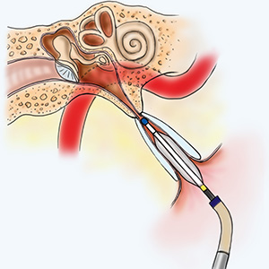
This simple procedure seems to restore normal ET function and reduce or resolve patient symptoms. Reported complications from balloon eustachian tuboplasty are relatively rare and mild.
Because the Eustachian tube is close to the internal carotid artery, there is concern about injury to this critical structure by inflating the balloon in the Eustachian tube. To reduce this risk, most surgeons will obtain a preoperative imaging study to confirm the anatomy of this region. In addition, specific devices have been designed to reduce the risk of ICA injury.