 Like a system of bayous, streams, and drains, the lymphatic system transports fluid throughout the body, and in the process, maintains fluid levels, removes waste, and elicits immune responses. As the bayous are essential to Houston during a rainstorm or hurricane, the lymphatic system is key to health and well-being.
For more than two decades, Eva Sevick-Muraca, PhD, director of the IMM’s Center for Molecular Imaging, and her team have literally shone a light on the lymphatics of more than 700 infants, children, and adults in the Texas Medical Center via innovative infrared fluorescent imaging.
“The lymphatics play really important roles in many diseases associated with aging, including peripheral vascular disease,” she said. “When people age, they get venous stasis ulcers, peripheral arterial disease, and diabetic foot ulcers. Through imaging, we’ve been able to show that the lymphatics play a role in the etiology of these conditions and could be an impactful target to treat and possibly even prevent them.”
The group also has used imaging for dosing therapeutics.
“Our work suggests that we can more effectively treat patients with autoimmune disease and cancer through regional lymphatic delivery as opposed to intravenous delivery of these drugs,” she said.
Colleague Melissa Aldrich, PhD, associate professor in the Center for Molecular Imaging, is using the lab’s imaging technology to evaluate breast cancer patients for lymphedema before surgery and every six months after radiation.
Through imaging, Aldrich and her collaborators at MD Anderson Cancer Center were able to see the onset of lymphatic abnormalities 8 to 23 months before the start of irreversible patient symptoms. “If we can treat at the first sign of lymphatic abnormalities, perhaps we can prevent its onset. I hope that with imaging diagnostics, lymphedema will be a disease of the past,” said Sevick, the Nancy and Rich Kinder Distinguished Chair of Cardiovascular Disease Research.
John Rasmussen, PhD, assistant professor in the Center for Molecular Imaging, specializes in engineering optical instrumentation and is currently adapting computer vision techniques to the lymphatic imaging technology that he co-invented. The additional technology will enable clinicians to longitudinally track lymphatic function in cancer survivors, providing an important clinical research tool for future clinical studies to prevent lymphedema.
Sevick and her team are turning their attention to the lymphatics of the brain. In collaboration with Manish Shah, MD, associate professor of pediatric neurosurgery and William J. Devane Distinguished Professor, they are looking at the role of lymphatics in pediatric traumatic brain injury.
“When you have a traumatic brain injury, such as a brain bleed often suffered by premature babies, hemoglobin within brain tissues can cause neuroinflammation. Normally, cerebrospinal fluid (CSF) drains into the lymphatics to remove brain waste. But if the lymphatics are impaired, then brain waste products cannot leave the brain, and the acute neuroinflammation becomes chronic,” Sevick explained.
Via an investigational drug study under review by the FDA, Sevick and her team will trace fluid flow in hydrocephalus patients by adding a safe, fluorescent dye to the infants’ CSF.
“We want to see where that CSF goes, or doesn’t go, and then devise more efficient strategies to reduce fluid and prevent neurological deficits,” Sevick said.
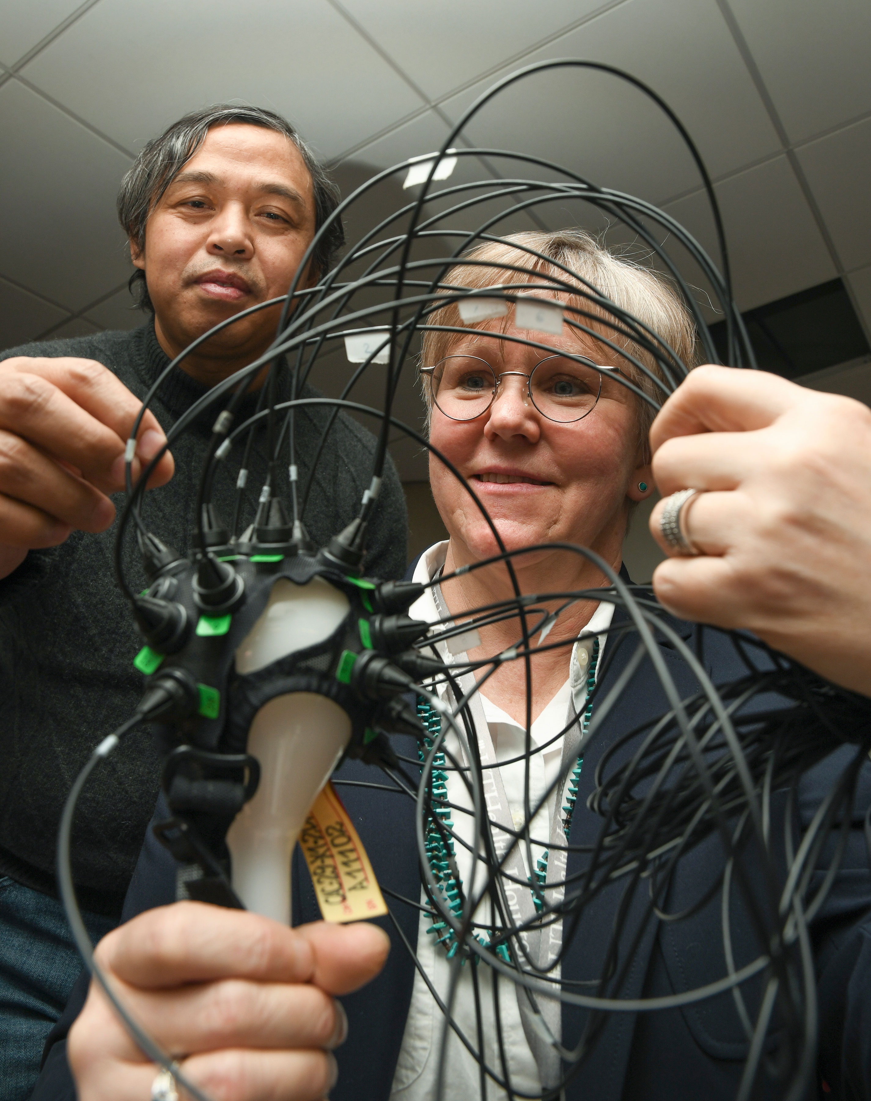
Banghe Zhu, PhD, assistant professor, Center for Molecular Imaging, engineered the specialized cap and optical imaging system, which generates 3D images.
While the team is currently focused on the pediatric population, studies to evaluate the lymphatic contribution are underway and planned in other neurodegenerative conditions.