Patient Information
PET and the Heart / Explanation of PET
Coronary Heart Disease
Coronary Heart Disease, the most common form of heart disease in this country and the primary cause of heart attacks, is the single largest killer of American men and women. Coronary Heart Disease occurs when the arteries carrying blood to the heart are blocked. Every 30 seconds, an American suffers a heart attack because of such a blockage, and 40 percent die within a year. More than 12 million Americans have been diagnosed with the disease and many more, an estimated 25% of the middle-aged population, are unaware of their illness until they succumb to an attack. In the U.S., coronary heart disease accounts for one-third of all deaths each year.
Positron Emission Tomography (PET) diagnoses heart disease non-invasively with 96-98% accuracy in individuals with or without symptoms of heart disease, permitting treatment even before symptoms appear. Furthermore, at our center, PET can help streamline efficient treatment of coronary heart disease, using quantitative coronary flow software to determine when invasive procedures such as stents or bypass surgery are absolutely necessary.
What is PET?
A cardiac PET scan is a type of stress test in which blood flow to the heart muscle is visualized with a radiotracer and a special scanner. PET provides an accurate non-invasive assessment of the coronary arteries. Pictures of the myocardial blood flow are taken before and after pharmacologic stress instead of exercise to determine if maximum blood flow is restricted by narrowing of the coronary arteries.
Why do a PET scan?
Since its diagnostic accuracy is much higher than standard tests at comparable cost per study, PET reduces expense and risk by avoiding unnecessary tests and procedures, thereby providing more efficient diagnoses. PET can be an alternative to invasive tests such as cardiac catheterization, in which a thin tube is threaded to the coronary arteries through an artery in the leg and X-ray pictures are taken after dye is injected into the heart. Even after an angiogram, PET can determine when a blockage is significant and a stent or coronary bypass surgery is necessary or whether medical management might be a better option.
There are 4 main reasons to have a cardiac PET scan:
- To be screened for early heart disease in patients who have no symptoms or have chest discomfort and have high cholesterol levels or a strong family history of heart disease.
- To check on the status of the coronary arteries in patients who have known coronary artery disease or who want to check on the progress of reversal treatment or other interventions (every 3-5 years or sooner if new symptoms develop).
- To determine the need for a coronary stent or coronary bypass surgery when blockages are found on an angiogram.
- To check on the viability of the heart muscle. If a large amount of heart tissue is scarred because of a heart attack, the heart will not pump well. The amount of damage and percentage of the heart affected will help doctors determine appropriate treatment including whether or not bypass surgery will be helpful.
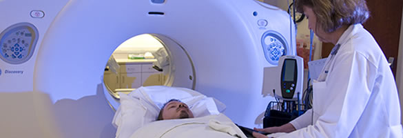
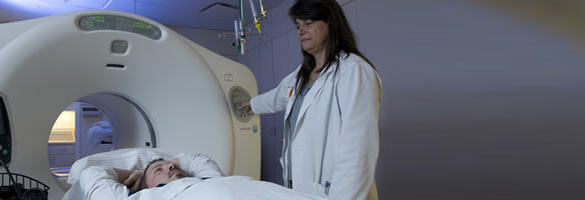
What will I experience at my PET scan appointment?
REST/STRESS IMAGING
After you have been checked in for your PET scan appt, you will be interviewed by a registered nurse to confirm your medical history, medications, and recent symptoms. The test will be explained in detail and you will sign a consent form. You will be brought to the PET preparation room to get your IV inserted and monitoring electrodes placed. Once started, the test should take about one hour.
The PET/CT camera is a large donut shaped camera that you will be placed in. You will not be enclosed (like an MRI) but you do have to move through a tunnel for about 2 minutes as we position you. Once in position, your head and arms will be outside the tunnel. You will be able to see the room and the people around you. If you think you are too claustrophobic to get through this, please ask your physician for a sedative medication to take before your scan.
We will inject a radioactive tracer to take the pictures of your heart muscle at rest and after a “chemical stress”. We do not use CONTRAST material to take the pictures so don’t worry about contrast allergies. You will not feel the radioactive tracer being injected to take the pictures but you may have symptoms with the stress medication. The stress medication brings lots of blood to the heart so it is a normal reaction to feel chest fullness and a flushed feeling. If you regularly have chest pain with physical activity, the stress drug may bring that on, so we will be monitoring you very closely and a cardiologist will be in the room during the stress. You just need to talk to us and let us know how you are feeling and if you feel uncomfortable at all. After the pictures are started, we will administer the antidote to the medication.
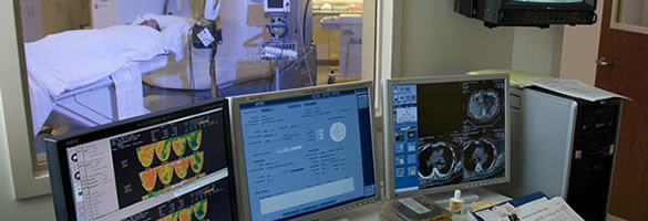
Patients referred by their private physician will be released after a few minutes and a full report of the results will be sent to their physician.
VIABILITY IMAGING
Sometimes, your physician may request VIABILITY imaging, especially if you have had a large heart attack in the past. If Viability imaging is required, it will add an additional 2-3 hours for your appt, but most of that time, you are sitting in a comfortable chair waiting.
After you have completed the Rest/stress part described above, you will be taken out of the camera, while we give you IV glucose and insulin to get your blood sugar to a certain level. When your blood sugar is right, we inject a radioactive sugar (FDG) to further investigate the scar in your heart. After the radioactive sugar has time to metabolize, you go back into the camera for a final scan.
I know this all sounds complicated, but we will always explain things and answer any questions you may have, as we go. Our goal is to make you as comfortable as possible and do the least amount of imaging necessary for you but also to make sure we get the information that your physician needs to take care of you
How do PET scans differ from other tests?
Clinical studies have shown that PET scans are more accurate than other widely used tests such as ECG (electrocardiogram), treadmill testing and standard nuclear stress tests such as SPECT (single photon emission computed tomography). These tests are associated with “false positive” and “false negative” results. False positives are results that show coronary heart disease where none really exists. False positives can lead to people undergoing unnecessary invasive procedures. Because PET scans are so accurate, they are often used to confirm other tests if a false positive or false negative is suspected. “False negatives” are normal results when coronary heart disease really exists. False negatives can lead to undetected heart disease.
Heart Scans (Fast CT) can detect the presence of calcium in the coronary arteries but can’t tell you if the calcium is affecting blood flow in the arteries. PET gives you both; information on the presence of calcium and an accurate quantitative measurement of the blood flow in the coronary arteries.
Medical Insurance Coverage
A PET Scan can be ordered by your physician and billed to insurance.
The ordering physician should send an order for the PET along with patient insurance information. The ordering physician who has the patient’s medical history should do pre-authorization. Orders should be faxed to 713-500-6615 and our office will contact the patient to schedule and review the financial coverage. A full report will be sent to the ordering physician. The Weatherhead PET Center has contracts with the following insurance companies:
- Blue Cross – HMO, PPO, Traditional
- United Healthcare
- Cigna (MedSolutions)
- Aetna
- Mhealth
- Medicare
- Humana
- Amerigroup
Our billing office will let you know if any payment is due before your appointment is scheduled. Patients will be responsible for their deductible and for the remainder of the charges not reimbursed by insurance. (Not to exceed the insurance allowable charge).