Atomic Force Microscopy Core Facility
Atomic Force Microscopy (external link) (AFM), an advanced multi-parametric imaging technique, not only delivers high-resolution 3D images of the topography of living biological samples, but also enables the characterization of the nanomechanical properties of molecules, cells, and tissues. AFM is an attractive tool for studying the systemic response to physiological processes. We can evaluate the stiffness of the sample surface by measuring the elastic properties (Young’s modulus) as a function of growth, differentiation, disease, or treatment. In addition, non-biological materials can also be analyzed.
Our core facility features the NanoWizard V AFM from JPK, Bruker Nano Inc., a state-of-the-art instrument that requires minimal sample preparation, making it ideal for delicate biological specimens. To enhance versatility, the NanoWizard V is integrated with a Nikon TE2000 inverted optical microscope, enabling simultaneous acquisition of bright-field and fluorescence images alongside AFM data. This powerful combination allows researchers to correlate structural, mechanical, and optical information in real time, providing a comprehensive view of sample properties.
Whether you are investigating cellular mechanics, biomolecular interactions, or material surfaces, our AFM platform delivers precision, reliability, and ease of use.
The services of the Atomic Force Microscopy Core include:
- Topographical imaging of samples in air or liquid environments
- High-resolution imaging at the nanometric scale
- Time-lapse experiments that show changes in sample morphology
- Nanoprobing of samples to quantify interaction forces
- Studies of local micromechanical properties (elasticity, stiffness, adhesion, roughness)
- Data analysis for assessing size distribution, peak force quantitative imaging, spectroscopy mapping and 3D imaging of a sample
(Services provided to internal/external users and the public sector)
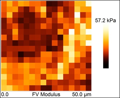
Spectroscopy map of a tissue sample
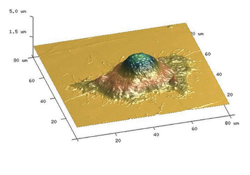
3D AFM image of a HeLa cell

3D AFM image of liposomes during internalization
 FOR FURTHER INFORMATION CONTACT:
ANA MARIA ZASKE, PhD
Research Scientist and Director of the AFM Core
UTHealth Houston
1881 East Road, 3SCRB 6.3728
Houston, Texas 77054
Phone: (713) 486-5418
E-mail: [email protected]