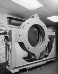
History
The PET Imaging Center was founded in 1979 by K. Lance Gould, M.D. as the Positron Diagnostic and Research Center at The University of Texas Health Science Center at Houston. Supported by a grant from the Clayton Foundation for Research, it developed into the world’s first major clinical PET Imaging Center. The results of the first 1,500 clinical heart studies at the PET Center indicated that PET was accurate in identifying early coronary heart disease as well as advanced, severe disease in people who had only mild or no symptoms, often years before a heart attack or the need for bypass surgery. But knowing the limitations of balloon dilation and bypass surgery, Dr. Gould sought a better way to treat patients that would not only identify early disease but also go a step further in preventing heart attacks and reversing the disease.

In the late 1980s and early 1990s, the Lifestyle Heart Trial with Dr. Dean Ornish demonstrated that lifestyle management improved blood flow in the hearts of patients with coronary heart disease. The PET scans in the study were done by Dr. Gould at the McGovern Medical School. In that study, PET imaging proved to be as accurate or more accurate than invasive coronary arteriograms for assessing the progression or regression of coronary heart disease.
Soon thereafter, a new class of cholesterol lowering drugs called statins was introduced and achieved a 30 to 75 percent decrease in the frequency of heart attacks, deaths, bypass surgery, or balloon dilation in patient with CAD. Good scientific data and subsequent clinical experience indicated that the combination of very low-fat, low calorie foods and cholesterol lowering drugs would provide the optimal treatment and maximal protection from heart attacks, balloon dilation, and bypass surgery. During this time, the technology of the PET camera itself continued to advance and a camera was put in at Memorial Hermann Hospital.
In December 1997, two of Dr. Gould’s patients, Al and Celia Weatherhead, recognized the great accomplishments and potential of PET imaging combined with reversal therapy, and pledged a generous 3 million dollar donation to assure the continued advancement of the PET Center. It then became the Weatherhead PET Center for Preventing and Reversing Atherosclerosis. A further donation by Mr. Weatherhead and other grateful patients funded the CENTURY Health Study; a 5-year randomized clinical trial of 1300 patients testing the use of PET and lifestyle management against standard cardiology care.
As the cardiology community began to recognize the limitations of standard cardiology treatments and the efficiency of treatment possible by using PET, the PET center has continued to evolve into a clinical imaging center with insurance contracts covering the costs of most scans. PET has become a tool to help determine when invasive stents or bypass surgery is absolutely necessary.
Most recently, the development of software to provide quantitative measurement of coronary blood flow continues to improve the utility of PET in the management of coronary heart disease.